. 2015 Sep;35(9):1987-94.
doi: 10.1161/ATVBAHA.115.305969. Epub 2015 Jul 9.

Orai3 Surface Accumulation and Calcium Entry Evoked by Vascular Endothelial Growth Factor

Affiliations

Orai3 Surface Accumulation and Calcium Entry Evoked by Vascular Endothelial Growth Factor

Jing Li et al. Arterioscler Thromb Vasc Biol. 2015 Sep.

Abstract

Objective: Vascular endothelial growth factor (VEGF) acts, in part, by triggering calcium ion (Ca(2+)) entry. Here, we sought understanding of a Synta66-resistant Ca(2+) entry pathway activated by VEGF.

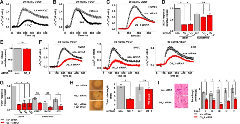
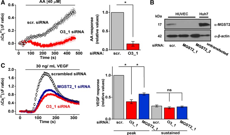
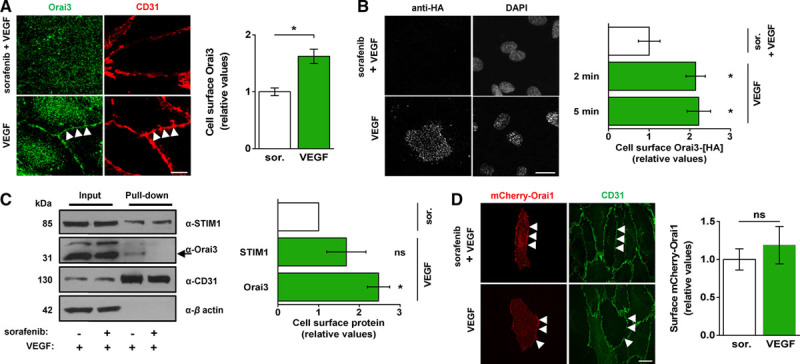
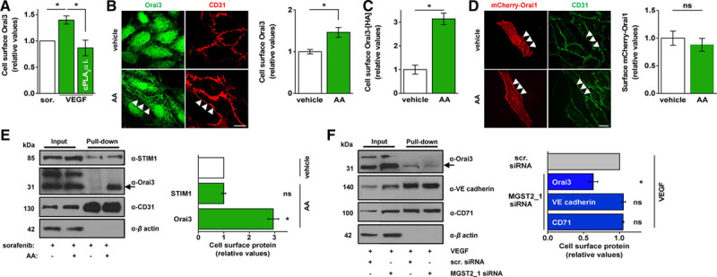
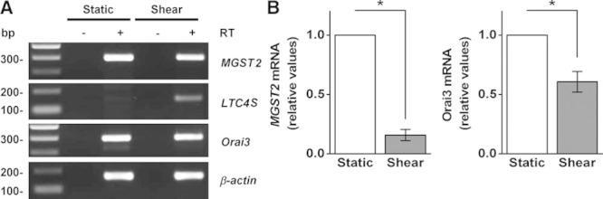
Approach and results: Measurement of intracellular Ca(2+) in human umbilical vein endothelial cells detected a Synta66-resistant component of VEGF-activated Ca(2+) entry that occurred within 2 minutes after VEGF exposure. Knockdown of the channel-forming protein Orai3 suppressed this Ca(2+) entry. Similar effects occurred in 3 further types of human endothelial cell. Orai3 knockdown was inhibitory for VEGF-dependent endothelial tube formation in Matrigel in vitro and in vivo in the mouse. Unexpectedly, immunofluorescence and biotinylation experiments showed that Orai3 was not at the surface membrane unless VEGF was applied, after which it accumulated in the membrane within 2 minutes. The signaling pathway coupling VEGF to the effect on Orai3 involved activation of phospholipase Cγ1, Ca(2+) release, cytosolic group IV phospholipase A2α, arachidonic acid production, and, in part, microsomal glutathione S-transferase 2, an enzyme which catalyses the formation of leukotriene C4 from arachidonic acid. Shear stress reduced microsomal glutathione S-transferase 2 expression while inducing expression of leukotriene C4 synthase, suggesting reciprocal regulation of leukotriene C4-synthesizing enzymes and greater role of microsomal glutathione S-transferase 2 in low shear stress.

Conclusions: VEGF signaling via arachidonic acid and arachidonic acid metabolism causes Orai3 to accumulate at the cell surface to mediate Ca(2+) entry and downstream endothelial cell remodeling.

Keywords: Orai3 protein; calcium; cytosol; endothelial cells; vascular endothelial growth factor A.


Figures

Figure 1.
Figure 1.
Role of Orai3 in vascular endothelial growth factor (VEGF)–evoked Ca2+ entry and endothelial cell remodeling. A, Example responses to VEGF stimulation (30 ng/mL) from human umbilical vein endothelial cells (HUVECs) in the presence or absence of Ca2+ (n=4/N=16). B, Example VEGF responses from HUVECs in the presence of Synta66 (S66; (5 μmol/L) or vehicle control (n=3/N=32 each). C, Example responses to VEGF stimulation (30 ng/mL) from HUVECs treated with scrambled (scr.) or Orai3_1 (O3_1) siRNA (n=3/N=60) each. D, Mean VEGF responses from HUVECs at peak or sustained (330 s) for cells transfected with scrambled (scr.), Orai3_1 (O3_1) or Orai3_2 (O3_2) siRNA (n=3/N=32 each). E, As for (C) but in the absence of extracellular Ca2+ (n=3/N=64 each). F, Example responses to VEGF stimulation (30 ng/mL) from cardiac microvascular endothelial cells (CMECs ; n=3/N=48 each), saphenous vein endothelial cells (SVECs; n=3/N=24 each), and liver endothelial cells (LECs; n=5/N=25 each). G, Mean data and analysis of data from CMECs (CM.), SVECs (SV.), and LECs as exemplified in (F). H, In vitro tube lengths of HUVECs after transfection with scr. or O3_1 siRNA; or scr. compared with O3_1 plus wild-type (WT) Orai3 clone (n=3 each). Scale bar, 50 μm. I, In vivo tube length of HUVECs in mice after transfection with scr. or O3_1 siRNA (5 independent experiments: i–v). Scale bars, 50 μm. Data are represented as mean±SEM. *P<0.05; not significant (ns) P>0.05.
Figure 2.
Figure 2.
Exogenous arachidonic acid (AA) evokes Orai3-dependent Ca2+ entry and microsomal glutathione S-transferase 2 (MGST2) is required in the action of vascular endothelial growth factor (VEGF). A, Example responses and mean data of human umbilical vein endothelial cells (HUVECs) exposed to exogenous AA (40 μmol/L). All cells were pretreated with thapsigargin (2 μmol/L) and studied in the presence of S66 (5 μmol/L). Cells were transfected with scrambled (scr.) or Orai3 (O3_1) siRNA; (n=3/N=60 each). B, Representative immunoblot of MGST2 in HUVECs transfected with scr. or MGST2 siRNA_1 or MGST2 siRNA_2. Untransfected Huh7 cells were used as a positive control for MGST2 expression. C, Example VEGF responses and mean data from HUVECs transfected with scr., O3_1 or MGST2_1 siRNA. Mean data are for VEGF responses at peak or after 330 s (n=3/N=36 each). Data are represented as mean±SEM; *P<0.05; ns P>0.05.
Figure 3.
Figure 3.
Selective Orai3 plasma membrane accumulation is evoked by vascular endothelial growth factor (VEGF). A, Representative images and mean data of cells treated with sorafenib (1 μmol/L) or vehicle (DMSO [dimethyl sulfoxide]) before stimulation with VEGF (30 ng/mL) for 5 minutes. Cells were labeled with anti-Orai3 antibody (Orai3, green) and anti-CD31 antibody (CD31, red). Scale bar, 2 μm. Arrows point to example cell perimeter as indicated by CD31 labeling. Mean data shows cell-surface Orai3 (n=6/N=18 each). B, Representative images and mean data of human umbilical vein endothelial cells (HUVECs) overexpressing Orai3-[HA] and treated as in (A). Cells were labeled with the anti-HA antibody. Scale bar, 10 μm. VEGF was applied for 5 minutes (n=3/N=15 each) or 2 minutes (n=3/N=45 each). C, Representative immunoblot and mean data from 3 experiments for cells treated as in (A) before biotinylation. The arrow points to Orai3 labeled by anti-Orai3 antibody (α-Orai3). The protein band above it, labeled nonspecifically by α-Orai3, has unknown identity. Where indicated (+), sorafenib (sor.) and VEGF were used at 1 μmol/L and 30 ng/mL, respectively. VEGF was applied for 5 minutes. D, Representative images and mean data for mCherry-Orai1 surface localization in cells treated as in (A); (sor., n=7/N=29; VEGF, n=4/N=14). Scale bar, 10 μm. All data are from HUVECs. Data are represented as mean±SEM; *P<0.05; ns P>0.05.
Figure 4.
Figure 4.
Selective Orai3 plasma membrane accumulation is evoked by exogenous arachidonic acid (AA) and depends on microsomal glutathione S-transferase 2 (MGST2). A, Mean immunofluorescence data for α-Orai3-labeled Orai3 in cells treated with sorafenib (1 μmol/L), vehicle or cytosolic group IV phospholipase A2α (cPLA2α) inhibitor (1 μmol/L) before stimulation with vascular endothelial growth factor (VEGF; 30 ng/mL) for 10 minutes (n=4/N=15 each). B, Representative images and mean data (n=4/N=22 each) of cells treated with sorafenib before stimulation with vehicle or AA (40 μmol/L) for 10 minutes. Cells were labeled with anti-Orai3 (green) and anti-CD31 (red) antibodies. Scale bar, 10 μm. Arrows point to cell perimeter as indicated by signal for CD31. C, Mean data for cells overexpressing Orai3-[HA] and treated as in (B; n=3/N=15 each). D, Representative images and mean data for mCherry-Orai1 surface localization in cells treated as in (B; vehicle control, n=7/N=29; AA, n=3/N=16 each). Scale bar, 10 μm. E, Representative immunoblot and mean data from 3 experiments for cell surface Orai3 from cells treated as in (B) before biotinylation. The arrow points to Orai3 labeled by anti-Orai3 antibody (α-Orai3). The protein band above it, labeled nonspecifically by α-Orai3, has unknown identity. Where indicated (+), sorafenib and AA were used at 1 μmol/L and 40 μmol/L, respectively. AA was applied for 5 minutes. F, Representative immunoblot and mean data from 3 experiments for cells treated with VEGF (30 ng/mL) for 5 minutes before biotinylation. The arrow points to Orai3 labeled by anti-Orai3 antibody (α-Orai3). The protein band above it, labeled nonspecifically by α-Orai3, has unknown identity. Where indicated (+), cells were transfected with control scrambled (scr.) siRNA or MGST2 siRNA_1. All data are from HUVECs. Data are represented as mean±SEM. STIM1 indicates stromal interaction molecule 1. *P<0.05; not significant (ns) P>0.05.
Figure 5.
Figure 5.
Analysis of MGST2, LTC4S, and Orai3 gene expression in human umbilical vein endothelial cell (HUVEC). A, Example agarose gel for end-point polymerase chain reaction products obtained with primers for MGST2, LTC4S, Orai3, and β-actin from HUVEC cDNA. Cells were exposed to static and shear-stress conditions, respectively, before harvesting mRNA and reverse transcriptase reaction (+RT [reverse transcriptase]) to generate cDNA. −RT denotes control reaction. B, Mean data and analysis of MGST2 and Orai3 mRNA (n=6). Data are represented as mean±SEM; *P<0.05.

References

    1. Feske S. ORAI1 and STIM1 deficiency in human and mice: roles of store-operated Ca2+ entry in the immune system and beyond. Immunol Rev. 2009;231:189–209. doi: 10.1111/j.1600-065X.2009.00818.x. - PMC - PubMed
    1. Cahalan MD. STIMulating store-operated Ca2+ entry. Nat Cell Biol. 2009;11:669–677. doi: 10.1038/ncb0609-669. - PMC - PubMed
    1. Lis A, Peinelt C, Beck A, Parvez S, Monteilh-Zoller M, Fleig A, Penner R. CRACM1, CRACM2, and CRACM3 are store-operated Ca2+ channels with distinct functional properties. Curr Biol. 2007;17:794–800. doi: 10.1016/j.cub.2007.03.065. - PMC - PubMed
    1. Motiani RK, Abdullaev IF, Trebak M. A novel native store-operated calcium channel encoded by Orai3: selective requirement of Orai3 versus Orai1 in estrogen receptor-positive versus estrogen receptor-negative breast cancer cells. J Biol Chem. 2010;285:19173–19183. doi: 10.1074/jbc.M110.102582. - PMC - PubMed
    1. Bisaillon JM, Motiani RK, Gonzalez-Cobos JC, Potier M, Halligan KE, Alzawahra WF, Barroso M, Singer HA, Jourd’heuil D, Trebak M. Essential role for STIM1/Orai1-mediated calcium influx in PDGF-induced smooth muscle migration. Am J Physiol Cell Physiol. 2010;298:C993–C1005. doi: 10.1152/ajpcell.00325.2009. - PMC - PubMed

Publication types

MeSH terms