

How are pluripotent cells captured in culture?
- PMID: 26161037
- PMCID: PMC4490168
- DOI: 10.1007/s12522-014-0199-8
How are pluripotent cells captured in culture?
Abstract
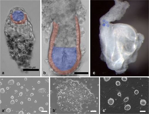
In mice, three pluripotent stem cell lines have been established from different stage of developing embryo, which are embryonic stem (ES) cell, post-implantation epiblast stem cell (EpiSC), and embryonic germ (EG) cell. ES cell and EG cell share many common features including factor requirement, colony morphology, and gene expression pattern. On the other hand, EpiSC needs different external signal inputs, exhibits flattened colony morphology, and a different set of gene expression patterns. In addition, the germ line competency of EpiSCs is still unclear. To distinguish the differences between them, they are defined by the words "naïve" and "primed" pluripotent cells, respectively. This article introduces how pluripotent stem cell lines are established in culture, and how much those cells in vitro are similar or relevant to their in vivo origin and the knowledge about transcription factors to support this state.
Keywords: EG cells; ES cell; EpiSC; Epiblast; Pluripotency.
Conflict of interest statement
Masaki Kinoshita declares that he has no conflicts of interest to disclose.
Figures
Similar articles
-
New cell lines from mouse epiblast share defining features with human embryonic stem cells.Nature. 2007 Jul 12;448(7150):196-9. doi: 10.1038/nature05972. Epub 2007 Jun 27. Nature. 2007. PMID: 17597760
-
Neural stem cells derived from epiblast stem cells display distinctive properties.Stem Cell Res. 2014 Mar;12(2):506-16. doi: 10.1016/j.scr.2013.12.012. Epub 2014 Jan 4. Stem Cell Res. 2014. PMID: 24463498
-
A Simplified and Efficient Protocol for Derivation and Maintenance of High-Quality Mouse Primed Pluripotent Stem Cells Using Wnt Inhibition.Curr Protoc Stem Cell Biol. 2018 Aug;46(1):e60. doi: 10.1002/cpsc.60. Epub 2018 Jul 13. Curr Protoc Stem Cell Biol. 2018. PMID: 30005143
-
Heterogeneity in Epiblast Stem Cells.Adv Exp Med Biol. 2019;1123:5-17. doi: 10.1007/978-3-030-11096-3_2. Adv Exp Med Biol. 2019. PMID: 31016592 Review.
-
Searching for naïve human pluripotent stem cells.World J Stem Cells. 2015 Apr 26;7(3):649-56. doi: 10.4252/wjsc.v7.i3.649. World J Stem Cells. 2015. PMID: 25914771 Free PMC article. Review.
Cited by
-
Generation of Naïve Bovine Induced Pluripotent Stem Cells Using PiggyBac Transposition of Doxycycline-Inducible Transcription Factors.PLoS One. 2015 Aug 19;10(8):e0135403. doi: 10.1371/journal.pone.0135403. eCollection 2015. PLoS One. 2015. PMID: 26287611 Free PMC article.
-
Derivation of Induced Trophoblast Cell Lines in Cattle by Doxycycline-Inducible piggyBac Vectors.PLoS One. 2016 Dec 1;11(12):e0167550. doi: 10.1371/journal.pone.0167550. eCollection 2016. PLoS One. 2016. PMID: 27907214 Free PMC article.
-
Trophoblast lineage specification in the mammalian preimplantation embryo.Reprod Med Biol. 2020 Jul 2;19(3):209-221. doi: 10.1002/rmb2.12333. eCollection 2020 Jul. Reprod Med Biol. 2020. PMID: 32684820 Free PMC article. Review.
References
-
- Yamanaka Y, Lanner F, Rossant J. FGF signal‐dependent segregation of primitive endoderm and epiblast in the mouse blastocyst. Development, 2010, 137, 715–724 - PubMed
-
- Nishioka N, Inoue K, Adachi K, Kiyonari H, Ota M, Ralston A et al. The Hippo signaling pathway components Lats and Yap pattern Tead4 activity to distinguish mouse trophectoderm from inner cell mass. Dev Cell, 2009, 16, 398–410 - PubMed
-
- Chazaud C, Yamanaka Y, Pawson T, Rossant J. Early lineage segregation between epiblast and primitive endoderm in mouse blastocysts through the Grb2‐MAPK pathway. Dev Cell, 2006, 10, 615–624 - PubMed
LinkOut - more resources
Full Text Sources
Other Literature Sources