. 2015:2015:412149.
doi: 10.1155/2015/412149. Epub 2015 Jun 16.

Sulforaphane Reverses the Expression of Various Tumor Suppressor Genes by Targeting DNMT3B and HDAC1 in Human Cervical Cancer Cells

Affiliations

Sulforaphane Reverses the Expression of Various Tumor Suppressor Genes by Targeting DNMT3B and HDAC1 in Human Cervical Cancer Cells

Munawwar Ali Khan et al. Evid Based Complement Alternat Med. 2015.

Abstract

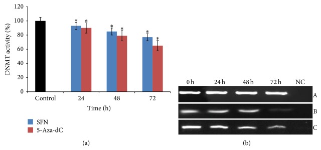
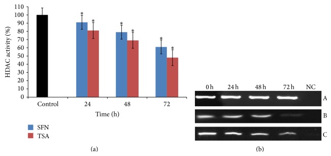
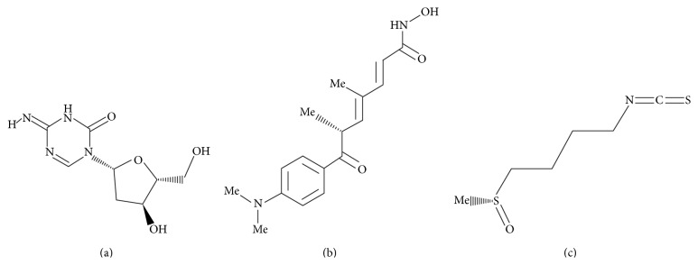
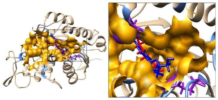
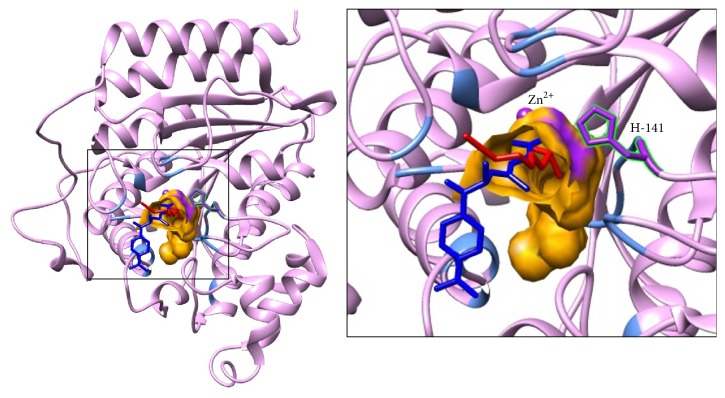
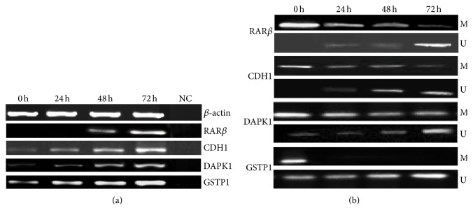
Sulforaphane (SFN) may hinder carcinogenesis by altering epigenetic events in the cells; however, its molecular mechanisms are unclear. The present study investigates the role of SFN in modifying epigenetic events in human cervical cancer cells, HeLa. HeLa cells were treated with SFN (2.5 µM) for a period of 0, 24, 48, and 72 hours for all experiments. After treatment, expressions of DNMT3B, HDAC1, RARβ, CDH1, DAPK1, and GSTP1 were studied using RT-PCR while promoter DNA methylation of tumor suppressor genes (TSGs) was studied using MS-PCR. Inhibition assays of DNA methyl transferases (DNMTs) and histone deacetylases (HDACs) were performed at varying time points. Molecular modeling and docking studies were performed to explore the possible interaction of SFN with HDAC1 and DNMT3B. Time-dependent exposure to SFN decreases the expression of DNMT3B and HDAC1 and significantly reduces the enzymatic activity of DNMTs and HDACs. Molecular modeling data suggests that SFN may interact directly with DNMT3B and HDAC1 which may explain the inhibitory action of SFN. Interestingly, time-dependent reactivation of the studied TSGs via reversal of methylation in SFN treated cells correlates well with its impact on the epigenetic alterations accumulated during cancer development. Thus, SFN may have significant implications for epigenetic based therapy.


Figures

Figure 1
Figure 1
Effect of SFN and 5-Aza-dC on DNMT3B in human cervical cancer cells (HeLa). (a) 2.5 μM of SFN and 1.5 μM 5-Aza-dC treatments significantly inhibit the activity of DNMT in time-dependent manner, respectively. Values are means ± SD of three independent experiments. Symbol () indicates significant (P < 0.05) difference of data between control and treated cells. (b) 2.5 μM SFN treated HeLa cells show significantly time-dependent reduction in the mRNA expression of DNMT3B in comparison to untreated cells. Panel A shows β-actin expression as an internal control, Panel B shows the expression of DNMT3B on treatment with 5-Aza-dC, and Panel C shows the expression of DNMT3B on treatment with SFN. Lane 1 shows the expression of DNMT3B gene in untreated HeLa cells; Lanes 2, 3, and 4 show the time-dependent alteration in the expression of DNMT3B after treatment for 24, 48, and 72 h, respectively; Lane 5 shows negative control for RT-PCR.
Figure 2
Figure 2
Effect of SFN and TSA on HDAC1 in human cervical cancer cells (HeLa). (a) 2.5 μM of SFN and 0.05 μM TSA treatments significantly inhibit the activity of HDAC in time-dependent manner, respectively. Values are means ± SD of three independent experiments. Symbol () indicates significant (P < 0.05) difference of data between control and treated cells. (b) 2.5 μM SFN treated HeLa cells show significantly time-dependent reduction in the mRNA expression of HDAC1 in comparison to untreated cells. Panel A shows β-actin expression as an internal control, Panel B shows the expression of HDAC1 on treatment with TSA, and Panel C shows the expression of HDAC1 on treatment with SFN. Lane 1 shows the expression of HDAC1 gene in untreated HeLa cells; Lanes 2, 3, and 4 show the time-dependent decrease in the expression of HDAC1 after treatment for 24, 48, and 72 h, respectively; Lane 5 shows negative control for RT-PCR.
Figure 3
Figure 3
Structures of the ligands used in the docking study. (a) 5-Aza-dC. (b) TSA. (c) SFN.
Figure 4
Figure 4
Predicted interaction between ligands (SFN and 5-Aza-dC) with mDNMT3B. The mDNMT3B is depicted in ribbon representation showing docked models of SFN in red and 5-Aza-dC in blue and the residues defining the pocket as light blue. Inset focuses on the binding pocket shown in orange. Active site C-651 and cofactor binding E-605 are labeled and shown in purple solid bonds.
Figure 5
Figure 5
Predicted interaction between ligands (SFN and TSA) with HDAC1. The HDAC1 protein is depicted in ribbon representation showing docked model of SFN in red and TSA in blue and the residues defining the pocket as light blue. The TSA structure was transformed from HDAC8 by superimposition on HDAC1. Inset focuses on the binding pocket shown in orange. The active site H-141 and Zn ion are labeled and highlighted in purple.
Figure 6
Figure 6
Alterations of methylation status and mRNA expression levels of RARβ, CDH1, DAPK1, and GSTP1 genes after treatment with SFN. (a) mRNA expression levels before and after the treatment. Lane 1 shows the expression of these genes in untreated HeLa cells; Lanes 2, 3, and 4 show the time-dependent modulation in the expression of HDAC1 upon treatment for 24, 48, and 72 h, respectively; Lane 5 shows negative control for RT-PCR. β-actin was used as an internal control. (b) Methylation-specific bands (M) and unmethylation-specific bands (U). Lane 1 shows the methylation status of these genes in untreated HeLa cells; Lanes 2, 3, and 4 show the time-dependent modulation in the methylation status of RARβ, CDH1, DAPK1, and GSTP1 genes for 24, 48, and 72 h, respectively.

Similar articles

Cited by

References

    1. You J. S., Jones P. A. Cancer genetics and epigenetics: two sides of the same coin? Cancer Cell. 2012;22(1):9–20. doi: 10.1016/j.ccr.2012.06.008. - DOI - PMC - PubMed
    1. Bilke S., Gindin Y. Evidence for a functional link between chromosomal breakpoints and aberrant DNA methylation in cancer. Frontiers in Oncology. 2014;4, article 46 doi: 10.3389/fonc.2014.00046.Article 46 - DOI - PMC - PubMed
    1. Virani S., Colacino J. A., Kim J. H., Rozek L. S. Cancer epigenetics: a brief review. ILAR Journal. 2012;53(3-4):359–369. doi: 10.1093/ilar.53.3-4.359. - DOI - PMC - PubMed
    1. Ivanova T., Petrenko A., Gritsko T., et al. Methylation and silencing of the retinoic acid receptor-beta 2 gene in cervical cancer. BMC Cancer. 2002;2(1, article 4) - PMC - PubMed
    1. Choi J. D., Lee J. S. Interplay between epigenetics and genetics in cancer. Genomics & Informatics. 2013;11(4):164–173. doi: 10.5808/gi.2013.11.4.164. - DOI - PMC - PubMed

LinkOut - more resources