. 2015 Jul 10;10(7):e0132503.
doi: 10.1371/journal.pone.0132503. eCollection 2015.

Hepatitis E Virus Produced from Cell Culture Has a Lipid Envelope

Affiliations

Hepatitis E Virus Produced from Cell Culture Has a Lipid Envelope

Ying Qi et al. PLoS One. .

Abstract

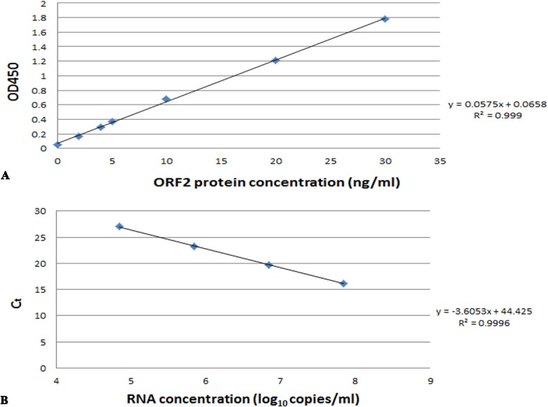
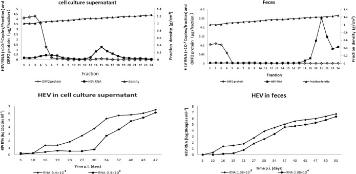
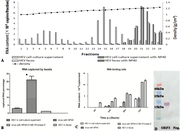
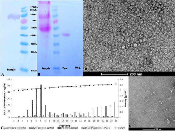
The absence of a productive cell culture system hampered detailed analysis of the structure and protein composition of the hepatitis E virion. In this study, hepatitis E virus from a robust HEV cell culture system and from the feces of infected monkeys at the peak of virus excretion was purified by ultra-centrifugation. The common feature of the two samples after ultracentrifugation was that the ORF2 protein mainly remained in the top fractions. The ORF2 protein from cell culture system was glycosylated, with an apparent molecular weight of 88 kDa, and was not infectious in PLC/PRF/5 cells. The ORF2 protein in this fraction can bind to and protect HEV RNA from digestion by RNase A. The RNA-ORF2 product has a similar sedimentation coefficient to the virus from feces. The viral RNA in the cell culture supernatant was mainly in the fraction of 1.15 g/cm3 but that from the feces was mainly in the fraction of 1.21 g/cm3. Both were infectious in PLC/PRF/5 cells. And the fraction in the middle of the gradient (1.06 g/cm3) from the cell culture supernatant,but not that from the feces, also has ORF2 protein and HEV RNA but was not infectious in PLC/PRF/5.The infectious RNA-rich fraction from the cell culture contained ORF3 protein and lipid but the corresponding fraction from feces had no lipid and little ORF3 protein. The lipid on the surface of the virus has no effect on its binding to cells but the ORF3 protein interferes with binding. The result suggests that most of the secreted ORF2 protein is not associated with HEV RNA and that hepatitis E virus produced in cell culture differs in structure from the virus found in feces in that it has a lipid envelope.


Conflict of interest statement

Competing Interests: The authors have declared that no competing interests exist.

Figures

Fig 1
Fig 1. The standard curve of ORF2protein (A) and HEV RNA (B).
(A): The X-axis is the concentration of ORF2 protein standard and Y-axis is the value of OD450. (B): X-axis is the concentration of HEV RNA standard and Y-axis is the Ct value of real time RT-PCR.
Fig 2
Fig 2. A and B are density-gradient fraction of HEV in cell culture supernatant (A) and feces (B).
The X-axis shows the fraction number, the left Y-axis is the ORF2 protein content (μg/ fraction) and the RNA content (105copies/fraction of cell culture supernatant HEV and 106 copies/fraction of feces HEV) in the fraction and the right Y-axis is the density of the fraction. C and D are quantification of HEV RNA in culture supernatant of PLC/PRF/5 cells incubated with virus from cell culture (C) and from feces (D). The X-axis is the time post incubation and the Y-axis is the HEV RNA concentration.
Fig 3
Fig 3. The analysis of the fraction rich inHEV RNA.
(A) The density-gradient fraction of virus from different sources and with different treatments. The X-axis is the fraction number and the right Y-axis is the density of the fraction. The left Y-axis is the HEV RNA content. (B) The binding ability of virus to anti-ORF3 MAb, the Y-axis is the percentage of binding HEV RNA to total HEV RNA of sample. The * represents the P value of HEV virus in the cell culture supernatant with NP40 treatment compared to the same sample without NP40 treatment. P value is less than 0.05. (C) The binding effect of virus to cells, the X-axis is the time post incubation with samples and the Y-axis was the binding RNA content to cells. (D) The fraction 15 of cell culture supernatant was detected by western blotting with an anti-ORF3 MAb.
Fig 4
Fig 4. Analysis of the ORF2 protein from the cell culture supernatant.
(A) Western blotting of the protein using an anti-ORF2 MAb. (B) The glycoprotein staining of the SDS-PAGE gel. The sample containsthe same magenta band as the positive control and the negative control has no band. (C) The RNA content in density-gradient fractions of the RNA-ORF2 binding product and controls. The X-axis is the fraction number, the left Y-axis is the HEV RNA content andthe right Y-axis is the density of the fraction.(D) and (E) Transmission electron microscopy of the RNA-ORF2 binding product (D) and ORF2 protein without HEV RNA(E) stained with uranyl acetate.The scale bar indicates 200 nm.
Fig 5
Fig 5. Specific Competitive Inhibition EMSA of HEV RNA and captured ORF2 protein.
The biotin-RNA without ORF2 protein was as negative control. The assay was carried out as described under “Materials and Methods”. The non-biotin labeled RNA in lane 2 and 3 were 4 fold and 8 fold to biotin labeled RNA. The lane 4 was shown the nonspecific competitive inhibition of 0.2μg total PLC/PRF/5 cell RNA.

References

    1. Aggarwal R, Naik S. Epidemiology of hepatitis E: current status. Journal of gastroenterology and hepatology. 2009;24(9):1484–93. Epub 2009/08/19. 10.1111/j.1440-1746.2009.05933.x . - DOI - PubMed
    1. Meng XJ. Zoonotic and foodborne transmission of hepatitis E virus. Seminars in liver disease. 2013;33(1):41–9. Epub 2013/04/09. 10.1055/s-0033-1338113 . - DOI - PubMed
    1. Agrawal S, Gupta D, Panda SK. The 3' end of hepatitis E virus (HEV) genome binds specifically to the viral RNA-dependent RNA polymerase (RdRp). Virology. 2001;282(1):87–101. Epub 2001/03/22. 10.1006/viro.2000.0819 . - DOI - PubMed
    1. Graff J, Torian U, Nguyen H, Emerson SU. A bicistronic subgenomic mRNA encodes both the ORF2 and ORF3 proteins of hepatitis E virus. Journal of virology. 2006;80(12):5919–26. Epub 2006/05/30. 10.1128/JVI.00046-06 - DOI - PMC - PubMed
    1. Yamada K, Takahashi M, Hoshino Y, Takahashi H, Ichiyama K, Nagashima S, et al. ORF3 protein of hepatitis E virus is essential for virion release from infected cells. The Journal of general virology. 2009;90(Pt 8):1880–91. 10.1099/vir.0.010561-0 . - DOI - PubMed

Publication types