. 2015 Jul 10;10(7):e0132637.
doi: 10.1371/journal.pone.0132637. eCollection 2015.

Caspase-Cleaved Tau Co-Localizes with Early Tangle Markers in the Human Vascular Dementia Brain

Affiliations

Caspase-Cleaved Tau Co-Localizes with Early Tangle Markers in the Human Vascular Dementia Brain

Ryan J Day et al. PLoS One. .

Abstract

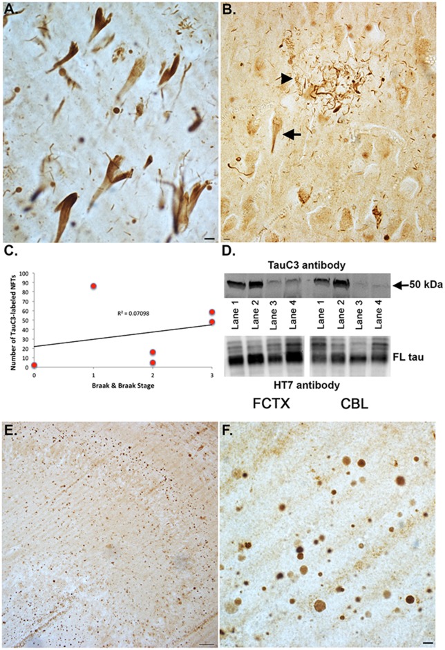
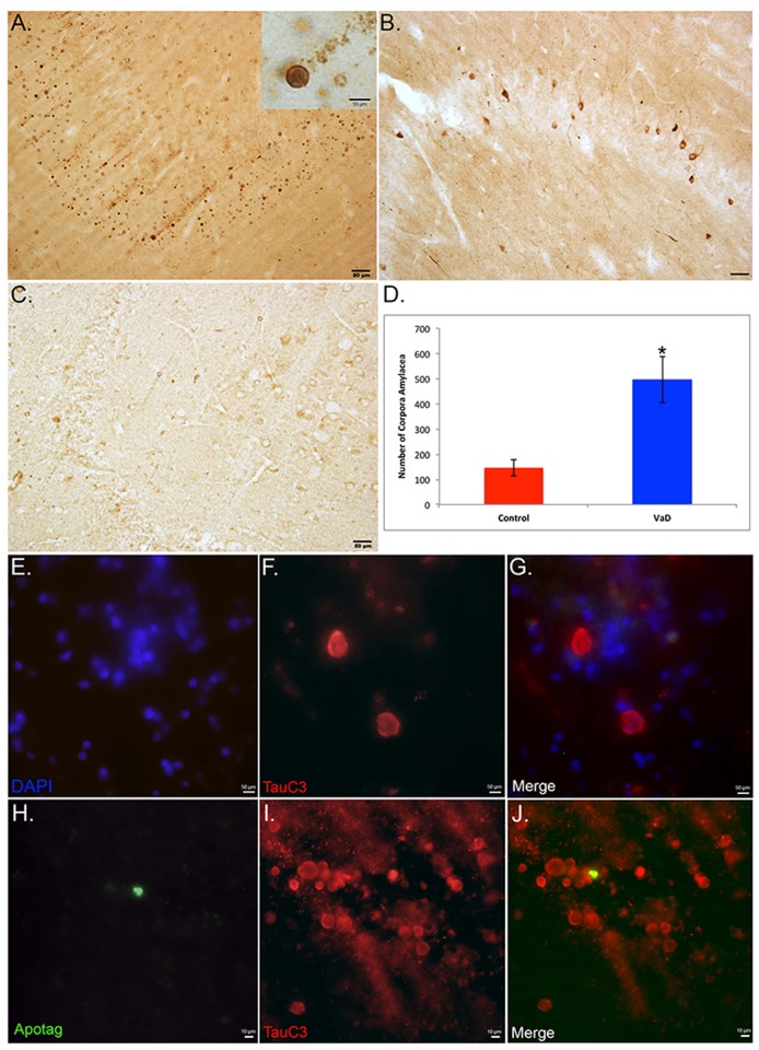
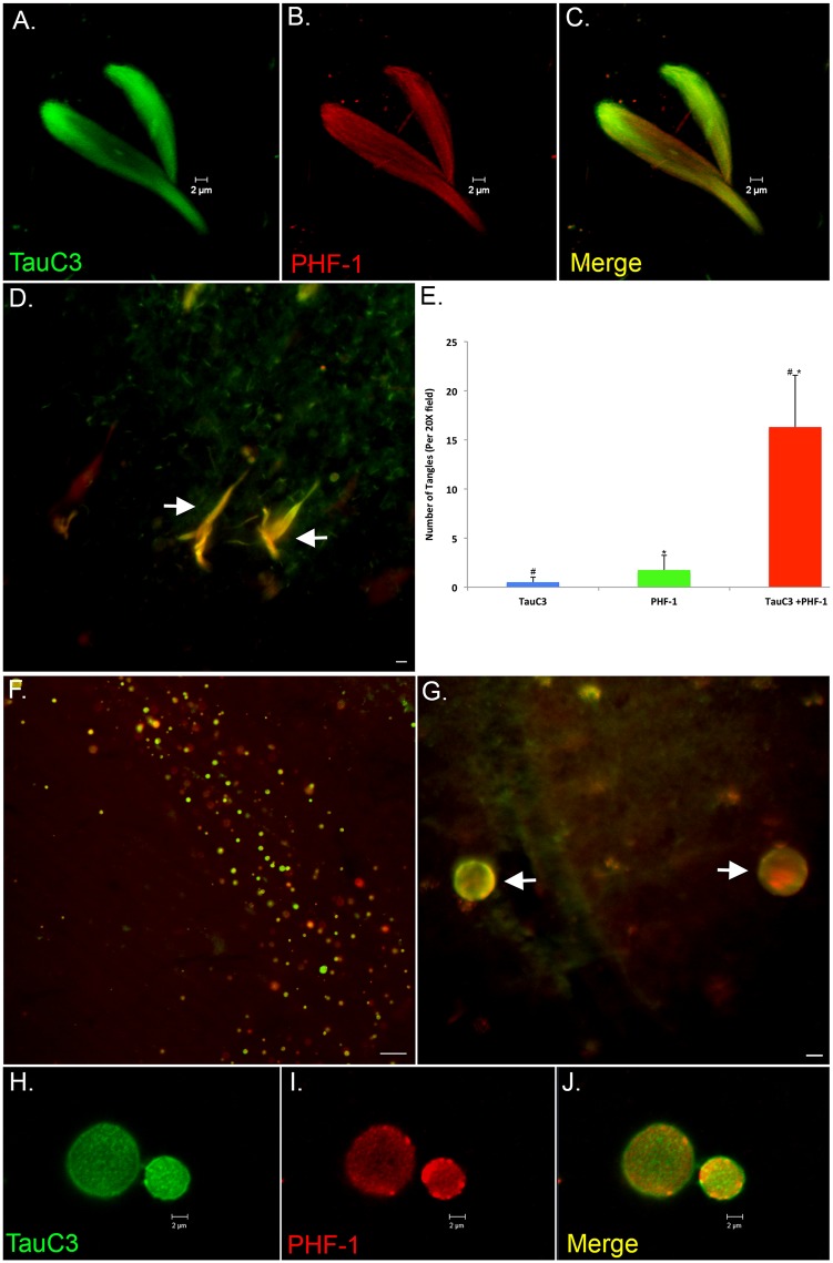
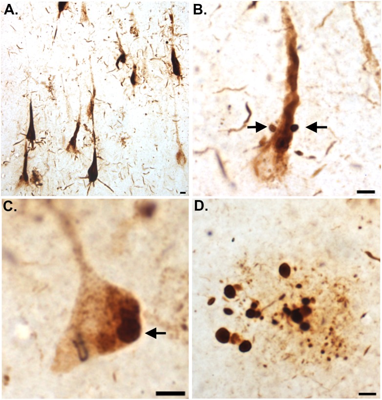
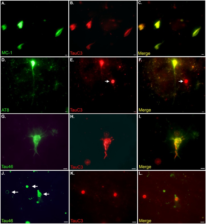
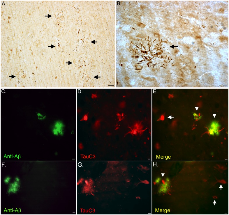
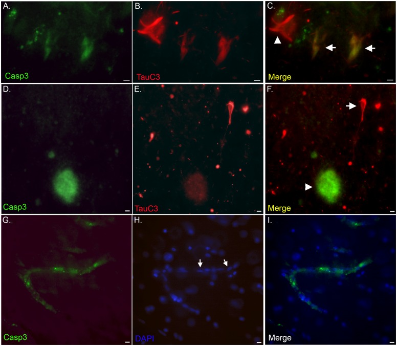
Vascular dementia (VaD) is the second most common form of dementia in the United States and is characterized as a cerebral vessel vascular disease that leads to ischemic episodes. Whereas the relationship between caspase-cleaved tau and neurofibrillary tangles (NFTs) in Alzheimer's disease (AD) has been previously described, whether caspase activation and cleavage of tau occurs in VaD is presently unknown. To investigate a potential role for caspase-cleaved tau in VaD, we analyzed seven confirmed cases of VaD by immunohistochemistry utilizing a well-characterized antibody that specifically detects caspase-cleaved tau truncated at Asp421. Application of this antibody (TauC3) revealed consistent labeling within NFTs, dystrophic neurites within plaque-rich regions and corpora amylacea (CA) in the human VaD brain. Labeling of CA by the TauC3 antibody was widespread throughout the hippocampus proper, was significantly higher compared to age matched controls, and co-localized with ubiquitin. Staining of the TauC3 antibody co-localized with MC-1, AT8, and PHF-1 within NFTs. Quantitative analysis indicated that roughly 90% of PHF-1-labeled NFTs contained caspase-cleaved tau. In addition, we documented the presence of active caspase-3 within plaques, blood vessels and pretangle neurons that co-localized with TauC3. Collectively, these data support a role for the activation of caspase-3 and proteolytic cleavage of TauC3 in VaD providing further support for the involvement of this family of proteases in NFT pathology.


Conflict of interest statement

Competing Interests: The authors have declared that no competing interests exist.

Figures

Fig 1
Fig 1. Caspase-cleaved tau in the human vascular dementia brain.
(A): Representative labeling from a VaD case utilizing the TauC3 antibody illustrating staining in the hippocampus within apparent NFTs. (B): Representative staining in the hippocampus indicating labeling of TauC3 within neuritic plaques (arrowhead), as well as apparent NFTs (arrow). (C): Correlation of TauC3 labeling with Braak & Braak stage. For six VaD cases in which the Braak & Braak stage was known, the number of TauC3-positive NFTs was counted three separate times, averaged and then plotted versus Braak & Braak stage. A positive correlation (R2 = .070) was observed between these two variables. (D): Western blot analysis utilizing the TauC3 antibody was carried out utilizing brain extracts from the frontal cortex (FCTX) or cerebellum (CBL) of four VaD cases. Lanes 1 (Case 6, Table 1) and 2 (Case 4, Table 1) are VaD cases that had Stage A plaque load, whereas lanes 3 (Case 3, Table 1) and 4 (Case 2, Table 1) were designated as having a plaque load of 0. A band at 50 kDa corresponding to caspase-cleaved tau truncated at Asp421 was identified in the FCTX of all four VaD cases and two of four cases in the CBL. The bottom panel of D depicts an identical experiment except transferred proteins were probed with HT7 (1:1,000), an antibody that detects total, full-length (FL) tau. (E and F): Low (E) and high magnification (F) of representative labeling from a VaD case utilizing the TauC3 antibody illustrating staining in the dentate gyrus of the hippocampus within numerous, round translucent structures. All scale bars represent 10 μm, except for Panel E, which represents 50 μm.
Fig 2
Fig 2. Identification of TauC3-labeled structures as apparent corpora amylacea in VaD.
(A): Bright-field staining utilizing the TauC3 antibody in the dentate gyrus of a representative VaD showing the presence of numerous round labeled structures that were ring-like in appearance (inset). (B): Representative bright-field staining utilizing HT7, an anti-body to full-length Tau did not label these round structures although numerous neurons were labeled. (C): Representative labeling of the TauC3 in an aged-matched control case indicating a relative paucity of labeling. Scale bars in Panels A-C represent 50 μm. (D): Quantitative analysis of the number of round structures in the hippocampi indicated a significant difference between VaD cases (n = 7, ±S.D.) and aged-matched controls (n = 4, ±S.D.), *p = .008. (E-G): Immunofluorescence double labeling in a representative VaD case utilizing TauC3 (red) and the nuclear stain, DAPI (blue) indicated that the round circular structures labeled by TauC3 are not nuclei (merge, G). (H-J): Immunofluorescence double labeling in a representative VaD case utilizing TauC3 (red) and TUNEL to label apoptotic cells (green) indicated that the round circular structures labeled by TauC3 are not apoptotic cells by in large (merge, J). Scale bars in Panels E-J represent 10 μm.
Fig 3
Fig 3. Confirmation of caspase-cleaved tau within corpora amylacea.
(A): Representative bright field staining in a VaD hippocampal brain section utilizing PAS that specifically labels CA in brain tissue. Labeled CA (magenta color, arrows) were in the vicinity of neurons in the granule cell layer of the dentate gyrus that were counter-stained with hematoxylin. (B): Representative bright-field labeling of numerous CA in the hippocampus of a VaD case utilizing an anti-ubiquitin, a specific marker for CA. (C and D): Representative immunofluorescence double-labeling in a VaD case at high magnification (C) and low magnification (D) indicating the co-localization of ubiquitin (green) together with the TauC3 antibody (red) within CA. In Panel D, the nuclei were also stained with nuclear stain, DAPI. All scale bars are equivalent to 10 μm except for Panel D, which represents 50 μm.
Fig 4
Fig 4. Co-localization of caspase-cleaved tau with NFTs in the VaD brain.
(A-C): Representative images from confocal immunofluorescence analysis in VaD utilizing TauC3 (green, A) and PHF-1 (red, B) with the overlap image shown in Panel C. Notice the filamentous nature of staining of PHF-1 as compared to TauC3. (D and E): Representative immunofluorescence double labeling (arrows, D) and quantification of NFTs (E) double-labeled with TauC3 and PHF-1. Data show the number of NFTs labeled with TauC3 alone (blue bar), PHF-1 alone (green bar) or NFTs that were labeled with both antibodies (red bar). NFTs were identified in a 20X field within hippocampi sections by immunofluorescence overlap microscopy (n = 3 fields for 4 different VaD cases) ±S.E.M. *p = 5.46 x 10⁻⁷ between PHF-1 alone and TauC3 + PHF-1 and #p = 4.49 x 10⁻⁷ between TauC3 alone and TauC3 + PHF-1. Data indicated that roughly 90% of all labeled NFTS co-localized with both antibodies. (F and G): Low- (F) and High-field (arrows, G) double immunofluorescence overlap images of corpora amylacea within the dentate gyrus of a representative VaD case showing co-localization of TauC3 (green) and PHF-1 (red). (H-J): High magnification confocal images representing labeling of corpora amylacea with TauC3 (H), PHF-1 (I), and the merged image (J). Scale bars represent 10 μm in Panels D and G and 50 μm for Panel F.
Fig 5
Fig 5. PHF-1 labeling of corpora amylacea within the hippocampus of the VaD brain.
(A): Representative bright-field DAB labeling from a VaD case utilizing the PHF-1 antibody illustrating staining in the hippocampus within NFTs. (B): High magnification of a single PHF-labeled NFT indicated the presence of small circular structures (arrows) of the same size and shape as CA in close proximity to the labeled neuron. (C): In this example, a PHF-labeled neuron appeared to exhibit intracellular inclusions that were of the same size and shape as identified CA (arrow). (D): Numerous CA labeled with PHF-1 were documented in plaque-rich regions within the hippocampal region of VaD cases. All scale bars represent 10 μm.
Fig 6
Fig 6. TauC3 co-localizes with early tangle markers in the VaD brain.
(A-C): Representative images from double-label immunofluorescence analysis in VaD utilizing the early pathological tau marker MC-1 (green, A) and TauC3 (red, B) with the overlap image shown in Panel C indicating co-localization of the two markers (yellow). (D-F): Representative double-label immunofluorescence experiment utilizing the early tangle phosphorylated marker, AT8 (green, D) and TauC3 (red, E) with the overlap image shown in Panel F. Strong co-localization between the two antibodies was observed in tangles, however, CA (arrow, E and F) labeled with TauC3 only. (G-I): Identical to Panels A-C except double-label was accomplished utilizing Tau46 (green, G), a C-terminal antibody to full-length tau. Note the distinct subcellular and fibrillary nature of the TauC3 antibody within the labeled neuron (H and I). (J-L): Identical to Panels G-I except the panels depict the labeling by Tau46 of CA (arrows, J) that were also labeled with TauC3 (K and L). All scale bars represent 10 μm.
Fig 7
Fig 7. Caspase-cleaved tau within neuritic plaques in the VaD brain.
(A and B): Representative bright-field microscopy showing TauC3 labeling of neuritic plaques at low-field (arrows, A) and at high magnification (arrow, B). (C-E): Representative immunofluorescence double labeling utilizing an anti-Aβ (clone 6E10) antibody (green, C), TauC3 (red, B), and the merged image (C) indicating co-localization of two markers (yellow, arrowheads). The arrow in Panel E reflects the labeling of a single NFT by TauC3 that is not labeled with the 6E10 antibody. (F-G): Identical to Panels C-E depicting labeling in an additional VaD case. All scale bars represent 10 μm except Panel A, which represents 50 μm.
Fig 8
Fig 8. Caspase-3 activation in the VaD brain.
(A-F): Representative immunofluorescence double labeling within the human VaD brain utilizing an antibody to active caspase-3 (green, Panels A and D) and TauC3 (red, Panels B and E), with the overlap images shown in Panels C and F. Labeling of active caspase-3 was evident within pretangles that co-localized with TauC3 (arrows, C). Co-localization of the two antibodies was also evident within plaques although TauC3 gave a much weaker fluorescence signal (F). In fibrillary NFTs only TauC3 was present (arrowhead, C and arrow, F). (G-I): Representative immunofluorescence double labeling with active caspase-3 (green, G) and the nuclear stain, DAPI (blue, H) indicating labeling within blood vessels of the VaD brain (I). Note the appearance of cuboid, elongated nuclei that typically define endothelial cell nuclei (arrows, H). All scale bars represent 10 μm.

Similar articles

Cited by

References

    1. Jellinger KA (2007) The enigma of vascular cognitive disorder and vascular dementia. Acta Neuropathol 113: 349–388. - PubMed
    1. Jellinger KA (2013) Pathology and pathogenesis of vascular cognitive impairment-a critical update. Front Aging Neurosci 5: 17 10.3389/fnagi.2013.00017 - DOI - PMC - PubMed
    1. Grinberg LT, Heinsen H (2010) Toward a pathological definition of vascular dementia. J Neurol Sci 299: 136–138. 10.1016/j.jns.2010.08.055 - DOI - PMC - PubMed
    1. Rohn TT (2014) Is apolipoprotein E4 an important risk factor for vascular dementia? Int J Clin Exp Pathol 7: 3504–3511. - PMC - PubMed
    1. Kalaria RN (2003) Comparison between Alzheimer's disease and vascular dementia: implications for treatment. Neurol Res 25: 661–664. - PubMed

Publication types