

Shear loads induce cellular damage in tendon fascicles
- PMID: 26162546
- PMCID: PMC5051692
- DOI: 10.1016/j.jbiomech.2015.06.006
Shear loads induce cellular damage in tendon fascicles
Abstract
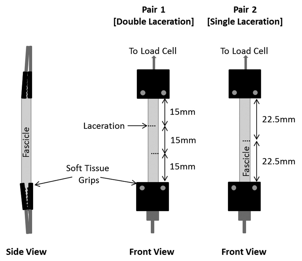
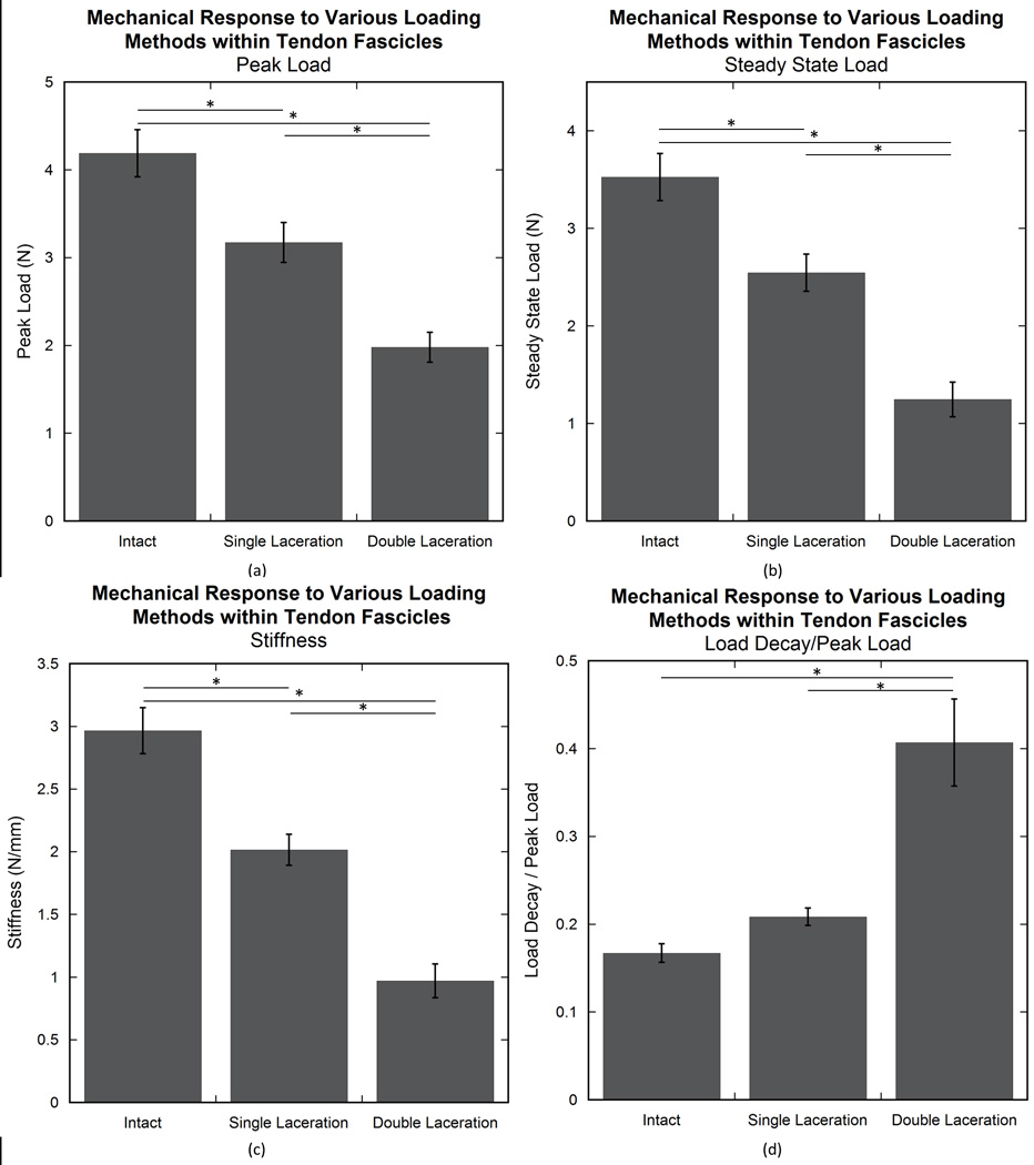
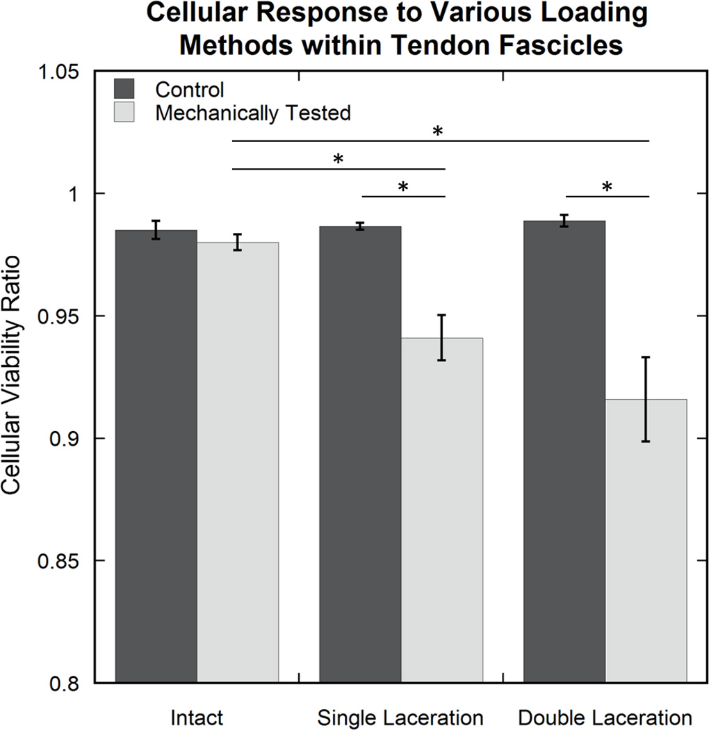
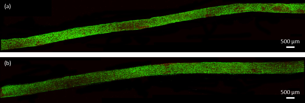
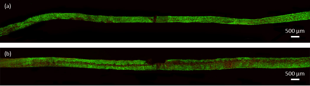
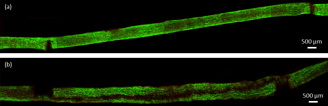
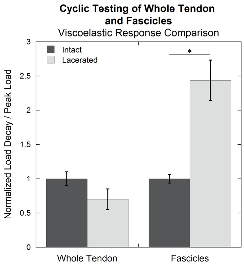
Tendon is vital to musculoskeletal function, transferring loads from muscle to bone for joint motion and stability. It is an anisotropic, highly organized, fibrous structure containing primarily type I collagen in addition to tenocytes and other extracellular matrix components contributing to maintenance and function. Tendon is generally loaded via normal stress in a longitudinal direction. However, certain situations, including fiber breakage, enzymatic remodeling, or tendon pathology may introduce various degrees of other loading modalities, such as shear-lag at the fiber level, potentially affecting cellular response and subsequent function. Fascicles from rat tail tendon were dissected and placed in one of three paired groups: intact, single laceration, or double laceration. Each pair had a mechanically tested and control specimen. Single laceration fascicles contained one transverse laceration to mimic a partial tear. Double laceration fascicles had overlapping, longitudinally separated lacerations on opposite sides to cause intra-fascicular shear transfer to be the primary mechanism of loading. Elastic properties of the fascicle, e.g. peak load, steady state load, and stiffness, decreased from intact to single laceration to double laceration groups. Surprisingly, 45% of the intact strength was maintained when shear was the primary internal load transfer mechanism. Cellular viability decreased after mechanical testing in both laceration groups; cell death appeared primarily in a longitudinal plane where high shear load transfer occurred. This cell death extended far from the injury site and may further compromise an already damaged tendon via enzymatic factors and subsequent remodeling associated with cell necrosis.
Keywords: Cellular viability; Mechanics; Shear; Tendon; Viscoelasticity.
Copyright © 2015 Elsevier Ltd. All rights reserved.
Conflict of interest statement
The authors have no conflict of interest.
Figures
Similar articles
-
Shear load transfer in high and low stress tendons.J Mech Behav Biomed Mater. 2015 May;45:109-20. doi: 10.1016/j.jmbbm.2015.01.021. Epub 2015 Feb 7. J Mech Behav Biomed Mater. 2015. PMID: 25700261 Free PMC article.
-
Tendon tissue microdamage and the limits of intrinsic repair.Matrix Biol. 2020 Jan;85-86:68-79. doi: 10.1016/j.matbio.2019.07.008. Epub 2019 Jul 17. Matrix Biol. 2020. PMID: 31325483
-
An in vitro scratch tendon tissue injury model: effects of high frequency low magnitude loading.Connect Tissue Res. 2017 Mar;58(2):162-171. doi: 10.1080/03008207.2016.1198338. Epub 2016 Jun 13. Connect Tissue Res. 2017. PMID: 27294971
-
Biomechanics and pathophysiology of overuse tendon injuries: ideas on insertional tendinopathy.Sports Med. 2004;34(14):1005-17. doi: 10.2165/00007256-200434140-00005. Sports Med. 2004. PMID: 15571430 Review.
-
Biology of tendon injury: healing, modeling and remodeling.J Musculoskelet Neuronal Interact. 2006 Apr-Jun;6(2):181-90. J Musculoskelet Neuronal Interact. 2006. PMID: 16849830 Review.
Cited by
-
Biplanar ultrasound investigation of in vivo Achilles tendon displacement non-uniformity.Transl Sports Med. 2019 Mar;2(2):73-81. doi: 10.1002/tsm2.61. Epub 2018 Dec 1. Transl Sports Med. 2019. PMID: 31008448 Free PMC article.
-
Interfibrillar shear behavior is altered in aging tendon fascicles.Biomech Model Mechanobiol. 2020 Jun;19(3):841-849. doi: 10.1007/s10237-019-01251-0. Epub 2019 Nov 9. Biomech Model Mechanobiol. 2020. PMID: 31707625 Free PMC article.
-
The Role of Detraining in Tendon Mechanobiology.Front Aging Neurosci. 2016 Feb 29;8:43. doi: 10.3389/fnagi.2016.00043. eCollection 2016. Front Aging Neurosci. 2016. PMID: 26973517 Free PMC article. Review.
-
Synergistic Biofilter Tube for Promoting Scarless Tendon Regeneration.Nano Lett. 2024 Jun 4;24(24):7381-8. doi: 10.1021/acs.nanolett.4c01540. Online ahead of print. Nano Lett. 2024. PMID: 38833276 Free PMC article.
-
Investigating mechanisms of tendon damage by measuring multi-scale recovery following tensile loading.Acta Biomater. 2017 Jul 15;57:363-372. doi: 10.1016/j.actbio.2017.04.011. Epub 2017 Apr 21. Acta Biomater. 2017. PMID: 28435080 Free PMC article.
References
-
- Abrahams M. Mechanical behaviour of tendon In vitro. Medical and Biological Engineering. 1967;5(5):433–443. http://doi.org/10.1007/BF02479137. - DOI - PubMed
-
- Ahmadzadeh H, Connizzo BK, Freedman BR, Soslowsky LJ, Shenoy VB. Determining the contribution of glycosaminoglycans to tendon mechanical properties with a modified shear-lag model. Journal of Biomechanics. 2013;46(14):2497–2503. http://doi.org/10.1016/j.jbiomech.2013.07.008. - DOI - PMC - PubMed
-
- Alberts B, Johnson A, Lewis J, Raff M, Roberts K, Walter P. Molecular Biology of the Cell. 4th. Garland Science; 2002.
-
- Banes AJ, Horesovsky G, Larson C, Tsuzaki M, Judex S, Archambault J, Miller L. Mechanical load stimulates expression of novel genesin vivoandin vitroin avian flexor tendon cells. Osteoarthritis and Cartilage. 1999;7(1):141–153. http://doi.org/10.1053/joca.1998.0169. - DOI - PubMed
-
- Bey MJ, Ramsey ML, Soslowsky LJ. Intratendinous strain fields of the supraspinatus tendon: Effect of a surgically created articular-surface rotator cuff tear. Journal of Shoulder and Elbow Surgery. 2002;11(6):562–569. - PubMed
Publication types
MeSH terms
Grants and funding
LinkOut - more resources
Full Text Sources
Other Literature Sources
