. 2015 Jul 28;112(30):9334-9.
doi: 10.1073/pnas.1503653112. Epub 2015 Jul 10.

Primer retention owing to the absence of RNase H1 is catastrophic for mitochondrial DNA replication

Affiliations

Primer retention owing to the absence of RNase H1 is catastrophic for mitochondrial DNA replication

J Bradley Holmes et al. Proc Natl Acad Sci U S A. .

Abstract

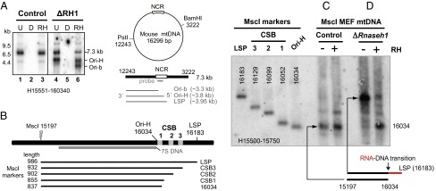
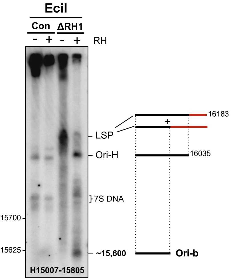
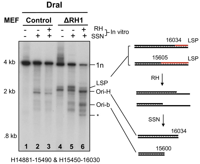
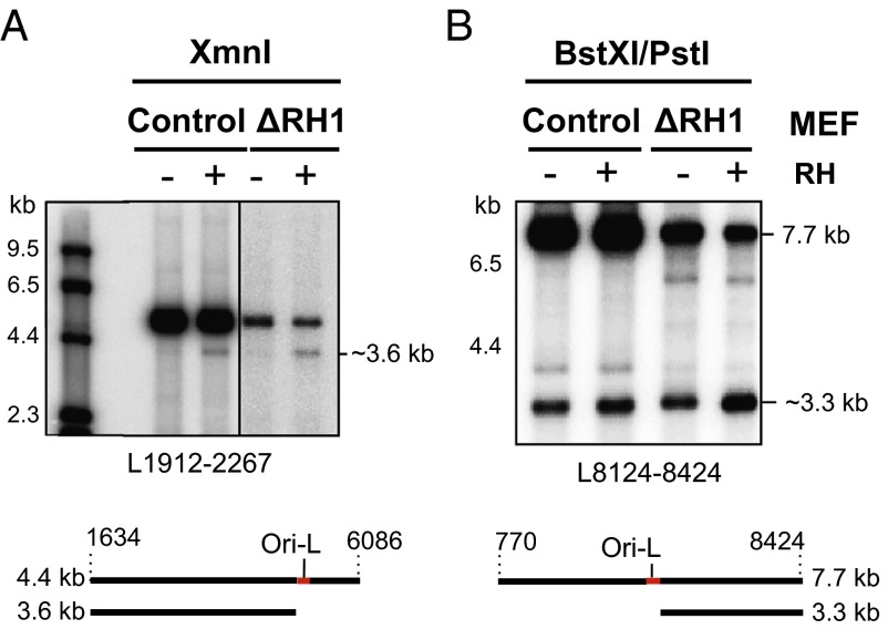
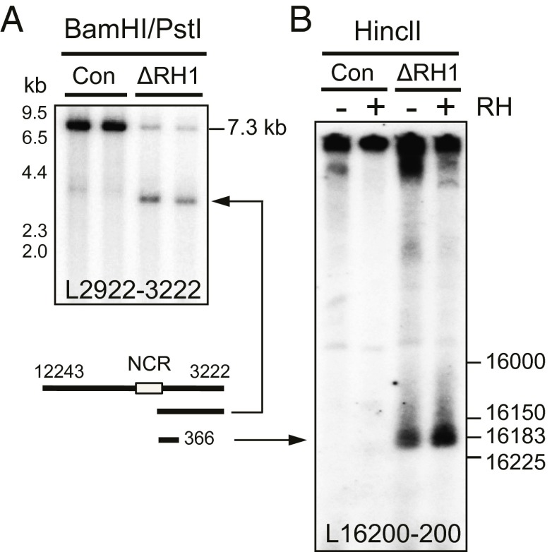
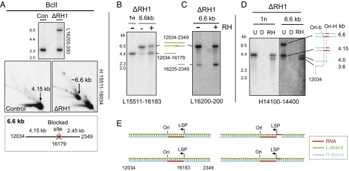
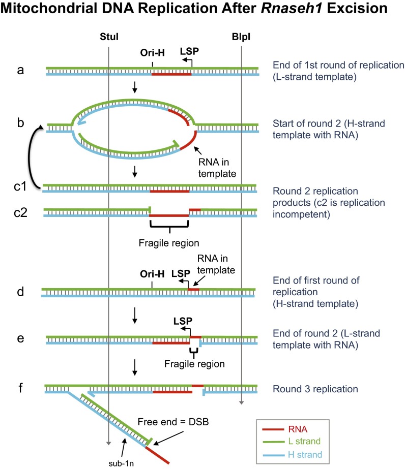
Encoding ribonuclease H1 (RNase H1) degrades RNA hybridized to DNA, and its function is essential for mitochondrial DNA maintenance in the developing mouse. Here we define the role of RNase H1 in mitochondrial DNA replication. Analysis of replicating mitochondrial DNA in embryonic fibroblasts lacking RNase H1 reveals retention of three primers in the major noncoding region (NCR) and one at the prominent lagging-strand initiation site termed Ori-L. Primer retention does not lead immediately to depletion, as the persistent RNA is fully incorporated in mitochondrial DNA. However, the retained primers present an obstacle to the mitochondrial DNA polymerase γ in subsequent rounds of replication and lead to the catastrophic generation of a double-strand break at the origin when the resulting gapped molecules are copied. Hence, the essential role of RNase H1 in mitochondrial DNA replication is the removal of primers at the origin of replication.

Keywords: DNA replication; RNase H; mitochondrial DNA; origin of replication; replication priming.


Conflict of interest statement

The authors declare no conflict of interest.

Figures

Fig. 1.
Fig. 1.
Loss of Rnaseh1 reveals two prominent origins of replication in the NCR and a primer starting at the light strand promoter. (A) MEF mtDNA, from cells treated for 9 d without (control) or with 4HT to excise the Rnaseh1 gene (ΔRH1), was digested with BamHI and PstI, treated with DNase (D) or Eco-RNase HI (RH) where indicated, denatured in 80% (vol/vol) formamide, 15 min at 85 °C, and separated by 1D-AGE (1% agarose Tris-acetate). λ-HindIII dsDNA ladder was run in parallel to provide size markers. Southern hybridization with a riboprobe to the 3′ end of the H-strand (H15551-160340) detected the full-length H-strand of 7.3 kb and one or two truncated species without or with Eco-RNase HI treatment (RH), respectively, whose lengths indicate their 5′ ends map to sites within the major NCR of mouse mtDNA (denoted LSP, Ori-H and Ori-b, see main text and B). (B) Map of the major NCR of murine mtDNA and the MscI digested markers and probe used for fine mapping of 5′ ends of DNA in the vicinity of Ori-H. MscI digested and denatured mtDNAs were fractionated by 1D-AGE and hybridized to riboprobe H15500-15750. Where indicated, samples were treated with (+) or without (−) Eco-RNase HI, before denaturation. DNA from mitochondria of MEFs treated without (C) or with 100 nM 4HT for 8 d (D). Interpretations of the RNase H sensitive species appear below C and D; red lines, RNA; black lines, DNA.
Fig. 2.
Fig. 2.
Rnaseh1 ablation leads to the accumulation of free 5′ ends of DNA mapping to nucleotide position ∼15,600 of the heavy strand of mouse mtDNA. EciI digested and denatured [80% (vol/vol) formamide, 15 min at 85 °C] MEF mtDNAs were fractionated by 1D-AGE and blot hybridized to riboprobe H15007-15805. Where indicated, samples were treated with (+) or without (−) Eco-RNase HI before denaturation. ΔRH1, mtDNA from MEFs treated with 100 nM 4HT for 8 d; Con, control MEFs. Interpretations of the single-stranded products are illustrated beside the gel images; red lines, RNA; black lines, DNA. Markers are shown in SI Appendix, Fig. S3.
Fig. 3.
Fig. 3.
Persistent RNA patches after Rnaseh1 ablation revealed by a combined RNase H and single-stranded nuclease treatments. (A) MEF mtDNA from cells culture for 7 d without (control) or with (ΔRH1) 4HT was digested with DraI, and where indicated additionally with single stranded nuclease (SSN) and Eco-RNase HI (RH), before native 1D-AGE, 1% agarose TAE. Fragments were blot hybridized to riboprobes H14881-15490 and H15450-16030. Interpretations of the products are illustrated beside the gel images; red lines, RNA; black lines, DNA. A truncated fragment mapping to nt ∼15,200 (*) was not reproducible (SI Appendix, Fig. S5I).
Fig. 4.
Fig. 4.
Rnaseh1 ablation leads to the accumulation of fully incorporated primers at Ori-L. MEF mtDNA from cells culture for 10 (A) or 9 (B) d without (control) or with (ΔRH1) 4HT was restriction digested with XmnI (A) or BstXI and PstI (B), denatured, and fractionated by 1D-AGE (1% agarose Tris-acetate). After transfer the fragments were hybridized with riboprobes (A) L1912-2267 or (B) L8124-8424. RH, samples were treated with (+) or without (−) Eco-RNase HI. (A) All samples were fractionated on the same gel, a longer exposure of the ΔRH1 samples is shown to compensate for unequal loading. (A and B) Below the gel images are interpretations of the RNase H sensitive species (a black line of DNA with a red RNA patch).
Fig. 5.
Fig. 5.
Loss of Rnaseh1 leads to retention of a RNA patch on the L-strand close to LSP. MEF mtDNA from cells cultured for 10 (A) or 8 (B) d with 4HT (ΔRH1) or from MEFs cultured without drug was digested with BamHI and PstI (A) or HincII (B), denatured for 15 min at 85 °C, in 80% (vol/vol) formamide, fractionated by 1D-AGE for 6 h at 11 V/cm, and blot hybridized to the indicated L-strand probe. RH, samples were treated with (+) or without (−) Eco-RNase HI. (A) 1% agarose Tris-acetate gel. (B) 2.3% (wt/vol) agarose sodium borate gel. Markers are based on PCR products of defined lengths (SI Appendix, Fig. S6).
Fig. 6.
Fig. 6.
Loss of Rnaseh1 leads to the retention of RNA patches on the H- and L-strands close to LSP, some of which are contiguous with DNA at both ends. BclI-digested mtDNA from MEFs cultured for 9 d without (control) or with (ΔRH1) 4HT was fractionated by (A) 2D-AGE or (B–D) 1D-AGE and blot hybridized to the indicated probes. For 1D-AGE, samples were denatured before loading. (Inset below A) Blocked BclI site at np 16,179 predicts a fragment of 6.6 kb. The BclI product of ∼6.6. kb was gel extracted, denatured, and refractionated after treatment without or with Eco-RNase HI (RH) or (D)Nase (B–D); next to the gel images are interpretations of the single-stranded products and (E) the possible double-stranded combinations, with Ori-H and Ori-b conflated to Ori. (B) Gel-extracted 4.15-kb fragment (1n) provided an additional marker, and in D, the same 1n material enabled comparison of the relative amounts of Ori-H and Ori-b ends of the 1n and 6.6-kb fragments.
Fig. 7.
Fig. 7.
Model of aberrant mitochondrial DNA replication in the absence of RNase H1. Primers on the H-strand from the LSP to the origin of replication Ori-H (or Ori-b, not illustrated) persist when RNase H1 is absent, and these are fully incorporated into mtDNA at the end of the replication cycle. In the next round of replication, the mitochondrial DNA polymerase γ (POLG) encounters a stretch of 150 (or 550) ribonucleotides of which it is expected to be able to synthesize only one or a few ribonucleotides, as it is a poor RNA-dependent DNA polymerase (29). RNase H1 ablation revealed a RNA patch on the L-strand near LSP that will prevent completion of H-strand synthesis once it is incorporated in the template strand. In the subsequent round of replication, the gap in the template H-strand effectively creates a double-strand break (DSB) at the origin (irrespective of RNA in the H-strand template).

References

    1. Bayona-Bafaluy MP, et al. Revisiting the mouse mitochondrial DNA sequence. Nucleic Acids Res. 2003;31(18):5349–5355. - PMC - PubMed
    1. Kasamatsu H, Vinograd J. Unidirectionality of replication in mouse mitochondrial DNA. Nat New Biol. 1973;241(108):103–105. - PubMed
    1. Chang DD, Hauswirth WW, Clayton DA. Replication priming and transcription initiate from precisely the same site in mouse mitochondrial DNA. EMBO J. 1985;4(6):1559–1567. - PMC - PubMed
    1. Crews S, Ojala D, Posakony J, Nishiguchi J, Attardi G. Nucleotide sequence of a region of human mitochondrial DNA containing the precisely identified origin of replication. Nature. 1979;277(5693):192–198. - PubMed
    1. Tapper DP, Clayton DA. Mechanism of replication of human mitochondrial DNA. Localization of the 5′ ends of nascent daughter strands. J Biol Chem. 1981;256(10):5109–5115. - PubMed

Publication types

LinkOut - more resources