

PSMA PET/CT with Glu-urea-Lys-(Ahx)-[⁶⁸Ga(HBED-CC)] versus 3D CT volumetric lymph node assessment in recurrent prostate cancer
- PMID: 26162799
- PMCID: PMC4589548
- DOI: 10.1007/s00259-015-3106-6
PSMA PET/CT with Glu-urea-Lys-(Ahx)-[⁶⁸Ga(HBED-CC)] versus 3D CT volumetric lymph node assessment in recurrent prostate cancer
Abstract
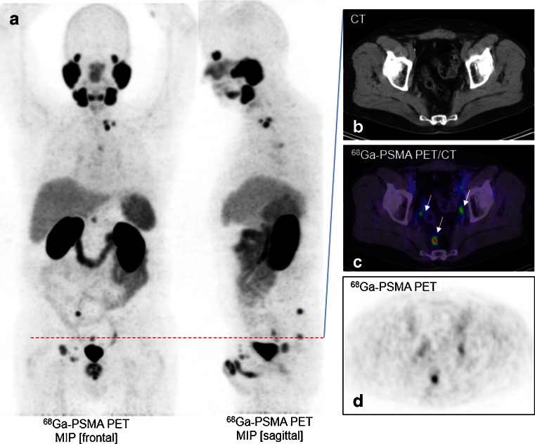
Purpose: PET/CT with the PSMA ligand is a powerful new method for the early detection of nodal metastases in patients with biochemical relapse. The purpose of this retrospective investigation was to evaluate the volume and dimensions of nodes identified by Glu-urea-Lys-(Ahx)-[(68)Ga(HBED-CC)] ((68)Ga-PSMA-11) in the setting of recurrent prostate cancer.
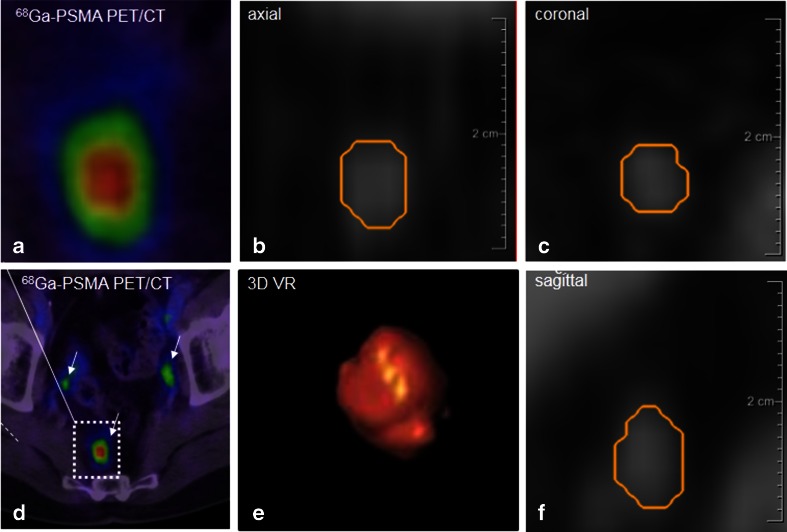
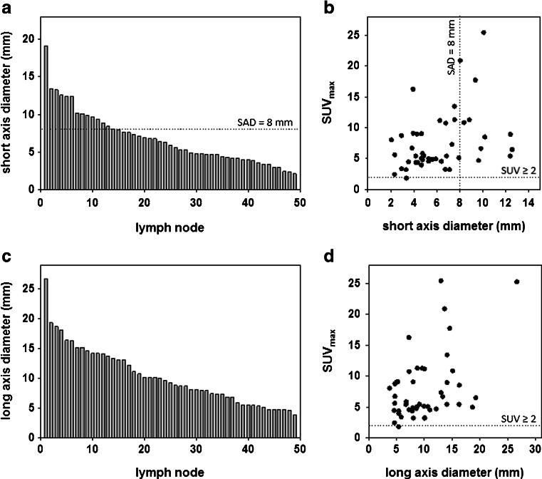
Methods: All PET/CT images were acquired 60 ± 10 min after intravenous injection of (68)Ga-PSMA-11 (mean dose 176 MBq). In 21 patients with recurrent prostate cancer and rising PSA, 49 PSMA-positive lymph nodes were identified. Using semiautomated lymph node segmentation software, node volume and short-axis and long-axis dimensions were measured and compared with the maximum standardized uptake values (SUVmax). Round nodes greater than or equal to 8 mm were considered positive by morphological criteria alone. The percentage of nodes identified by elevated SUVmax but not by conventional morphological criteria was determined.
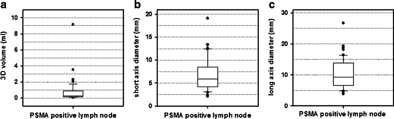
Results: The mean volume of (68)Ga-PSMA-11-positive nodes was 0.5 ml (range 0.2 - 2.3 ml), and the mean short-axis diameter was 5.8 mm (range 2.4 - 13.3 mm). In 7 patients (33.3 %) with 31 PSMA-positive nodes only 11 (36 %) were morphologically positive based on diameters >8 mm on CT. In the remaining 14 patients (66.7 %), 18 (37 %) of PSMA positive lymph nodes had short-axis diameters <8 mm with a mean short-axis diameter of 5.0 mm (range 2.4 - 7.9 mm). Thus, in this population, (68)Ga-PSMA-11 PET/CT detected nodal recurrence in two-thirds of patients who would have been missed using conventional morphological criteria.
Conclusion: (68)Ga-PSMA-11 PET/CT is more sensitive than CT based 3D volumetric lymph node evaluation in determining the node status of patients with recurrent prostate cancer, and is a promising method of restaging prostate cancers in this setting.
Keywords: 68Ga-PSMA-11 PET/CT; Lymph node evaluation; Lymph node metastasis; Recurrent prostate cancer.
Figures
Comment in
-
(68)Ga-PSMA-HBED-CC PET/CT: where molecular imaging has an edge over morphological imaging.Eur J Nucl Med Mol Imaging. 2016 Mar;43(3):394-6. doi: 10.1007/s00259-015-3212-5. Eur J Nucl Med Mol Imaging. 2016. PMID: 26452581 No abstract available.
References
Publication types
MeSH terms
Substances
LinkOut - more resources
Full Text Sources
Other Literature Sources
Medical
Research Materials
Miscellaneous
