

Virchow-Robin Spaces: Correlations with Polysomnography-Derived Sleep Parameters
- PMID: 26163465
- PMCID: PMC4434551
- DOI: 10.5665/sleep.4726
Virchow-Robin Spaces: Correlations with Polysomnography-Derived Sleep Parameters
Abstract
Study objectives: To test the hypothesis that enlarged Virchow-Robin space volumes (VRS) are associated with objective measures of poor quality sleep.
Design: Retrospective cross-sectional study.
Setting: Sunnybrook Health Sciences Centre.
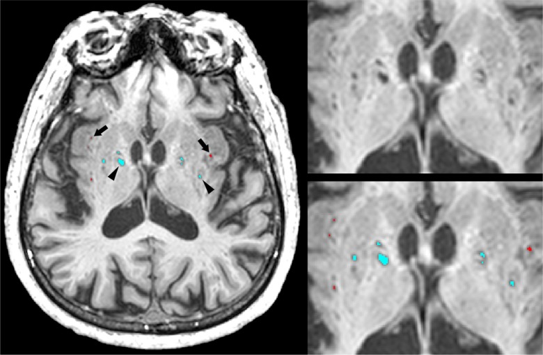
Patients: Twenty-six patients being evaluated for cerebrovascular disease were assessed using polysomnography and high-resolution structural magnetic resonance imaging.
Measurements and results: Regionalized VRS were quantified from three-dimensional high-resolution magnetic resonance imaging and correlated with measures of polysomnography-derived sleep parameters while controlling for age, stroke volume, body mass index, systolic blood pressure, and ventricular cerebrospinal fluid volume. Sleep efficiency was negatively correlated with total VRS (rho = -0.47, P = 0.03) and basal ganglia VRS (rho = -0.54, P = 0.01), whereas wake after sleep onset was positively correlated with basal ganglia VRS (rho = 0.52, P = 0.02). Furthermore, VRS in the basal ganglia were negatively correlated with duration of N3 (rho = -0.53, P = 0.01).
Conclusions: These preliminary results suggest that sleep may play a role in perivascular clearance in ischemic brain disease, and invite future research into the potential relevance of Virchow-Robin spaces as an imaging biomarker for nocturnal metabolite clearance.
Keywords: MRI; Virchow-Robin; basal ganglia; metabolite clearance; perivascular space; polysomnography; sleep; small vessel disease; stroke; white matter.
© 2015 Associated Professional Sleep Societies, LLC.
Figures
Comment in
-
Waking Up MRI-Visible Perivascular Spaces and Drainage Research.Sleep. 2015 Jun 1;38(6):845-6. doi: 10.5665/sleep.4718. Sleep. 2015. PMID: 26039964 Free PMC article. No abstract available.
References
-
- Abbott NJ. Evidence for bulk flow of brain interstitial fluid: significance for physiology and pathology. Neurochem Int. 2004;45:545–52. - PubMed
-
- Zhu YC, Tzourio C, Soumare A, Mazoyer B, Dufouil C, Chabriat H. Severity of dilated Virchow-Robin spaces is associated with age, blood pressure, and MRI markers of small vessel disease: a population-based study. Stroke. 2010;41:2483–90. - PubMed
-
- Rouhl RP, van Oostenbrugge RJ, Knottnerus IL, Staals JE, Lodder J. Virchow-Robin spaces relate to cerebral small vessel disease severity. J Neurol. 2008;255:692–6. - PubMed
Publication types
MeSH terms
Grants and funding
LinkOut - more resources
Full Text Sources
Other Literature Sources
Medical
