. 2015 Sep 15;309(6):L593-604.
doi: 10.1152/ajplung.00029.2015. Epub 2015 Jul 10.

Activation of the nuclear factor-κB pathway during postnatal lung inflammation preserves alveolarization by suppressing macrophage inflammatory protein-2

Affiliations

Activation of the nuclear factor-κB pathway during postnatal lung inflammation preserves alveolarization by suppressing macrophage inflammatory protein-2

Yanli Hou et al. Am J Physiol Lung Cell Mol Physiol. .

Abstract

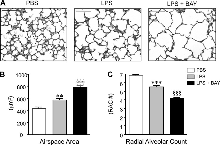
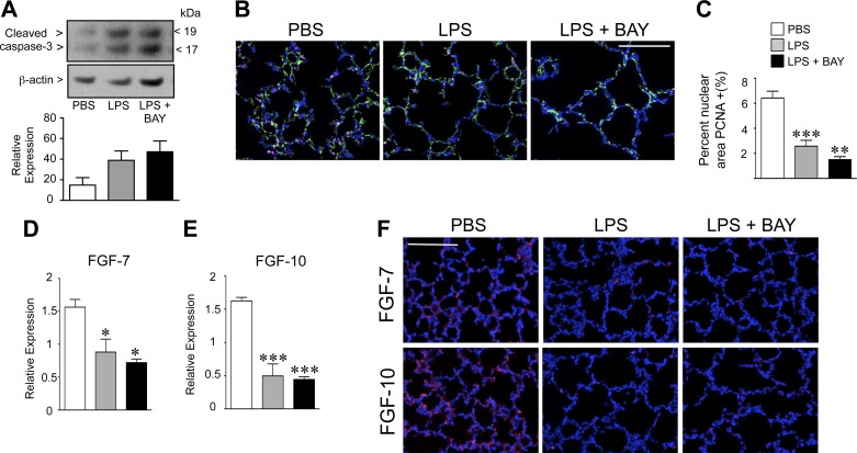
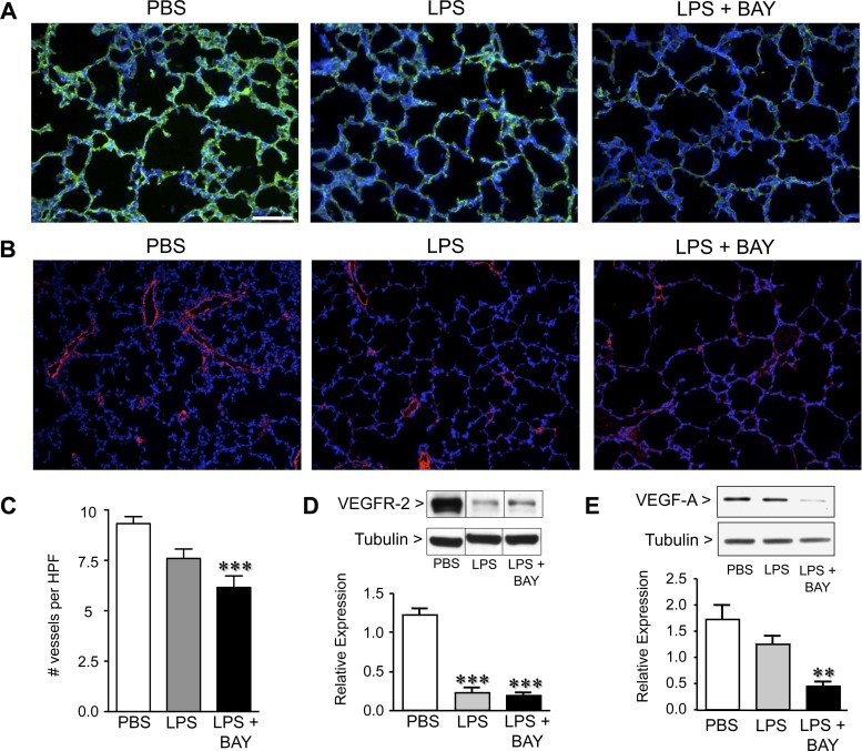
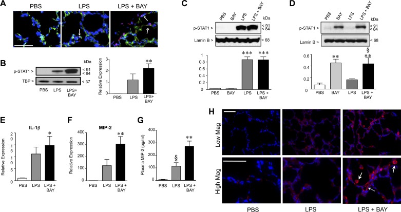
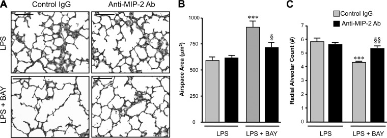
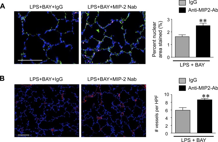
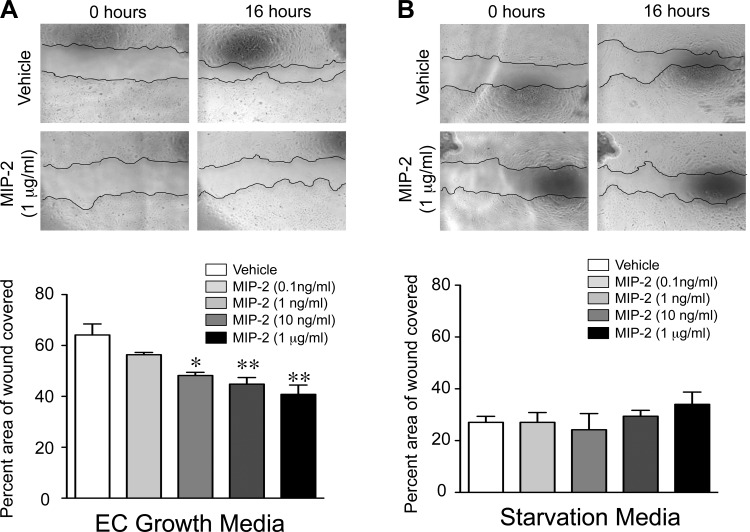
A significant portion of lung development is completed postnatally during alveolarization, rendering the immature lung vulnerable to inflammatory stimuli that can disrupt lung structure and function. Although the NF-κB pathway has well-recognized pro-inflammatory functions, novel anti-inflammatory and developmental roles for NF-κB have recently been described. Thus, to determine how NF-κB modulates alveolarization during inflammation, we exposed postnatal day 6 mice to vehicle (PBS), systemic lipopolysaccharide (LPS), or the combination of LPS and the global NF-κB pathway inhibitor BAY 11-7082 (LPS + BAY). LPS impaired alveolarization, decreased lung cell proliferation, and reduced epithelial growth factor expression. BAY exaggerated these detrimental effects of LPS, further suppressing proliferation and disrupting pulmonary angiogenesis, an essential component of alveolarization. The more severe pathology induced by LPS + BAY was associated with marked increases in lung and plasma levels of macrophage inflammatory protein-2 (MIP-2). Experiments using primary neonatal pulmonary endothelial cells (PEC) demonstrated that MIP-2 directly impaired neonatal PEC migration in vitro; and neutralization of MIP-2 in vivo preserved lung cell proliferation and pulmonary angiogenesis and prevented the more severe alveolar disruption induced by the combined treatment of LPS + BAY. Taken together, these studies demonstrate a key anti-inflammatory function of the NF-κB pathway in the early alveolar lung that functions to mitigate the detrimental effects of inflammation on pulmonary angiogenesis and alveolarization. Furthermore, these data suggest that neutralization of MIP-2 may represent a novel therapeutic target that could be beneficial in preserving lung growth in premature infants exposed to inflammatory stress.

Keywords: angiogenesis; bronchopulmonary dysplasia; endothelial migration; proliferation.


Figures

Fig. 1.
Fig. 1.
Systemic lipopolysaccharide (LPS) disrupts distal lung growth in the early alveolar lung, and inhibiting the NF-κB pathway accentuates these detrimental effects. A: representative images of hematoxylin and eosin (H&E)-stained lung sections obtained from postnatal day 7 (P7) mice, 24 h after administration of vehicle (PBS), LPS, or LPS + BAY 11-7082 (LPS + BAY). B: morphometric analysis to determine the distal airspace area, with **P < 0.01 vs. PBS and §§§P < 0.001 vs. PBS and LPS. C: morphometric analysis to determine the radial alveolar count, with ***P < 0.001 vs. PBS and §§§P < 0.001 vs. PBS and LPS. All data are expressed as means ± SE, with n = 6–9 animals per group. Scale bar = 100 μm. RAC, radial alveolar count.
Fig. 2.
Fig. 2.
Systemic LPS reduces proliferation and decreases epithelial growth factors in the early alveolar lung. A: Western immunoblot to detect cleaved-caspase 3 in lung tissue from PBS, LPS, and LPS + BAY-treated mice at 8 h. Data are expressed as means ± SE, with n = 4 to 5 for each group. B: representative composite immunofluorescent images from lung frozen sections obtained from PBS, LPS, and LPS + BAY-treated neonatal mice at 24 h, to detect p65 (red), CD31 (green), and chromatin (blue). Scale bar = 100 μm. C: quantification of the proliferating cell nuclear antigen (PCNA) positive nuclear area in the lungs of each group, with **P < 0.01 and ***P < 0.001 vs. PBS. Data are expressed as means ± SE, with n = 5 to 6 for each group. D: quantitative RT-PCR performed on whole lung, 8 h after either administration of PBS, LPS, or LPS + BAY to detect fibroblast growth factor (FGF)-7 and FGF-10 (E), with *P < 0.05 and ***P < 0.001 vs. PBS. Data are expressed as means ± SE, with n = 4 for each group. F: representative composite immunofluorescent images from lung frozen sections obtained from PBS, LPS, and LPS + BAY-treated neonatal mice at 24 h to detect either FGF-7 or FGF-10 (red) and chromatin (blue). Scale bar = 100 μm.
Fig. 3.
Fig. 3.
Systemic LPS disrupts pulmonary angiogenesis and decreases the expression of angiogenic factors, and inhibiting the NF-κB pathway accentuates these effects. A: representative immunofluorescent images from lung frozen sections obtained from PBS, LPS, and LPS + BAY-treated mice at 24 h, to detect CD31 (green) and chromatin (blue) (A) or von Willebrand factor (vWF; red) and chromatin (blue) (B). C: quantification of vWF-stained vessels per high power field (HPF) in each group with ***P < 0.001 vs. PBS. Data are expressed as means ± SE, with n = 5 to 6 for each group. D: Western immunoblot to detect VEGFR-2 and VEGF-A (E) in lung tissue from PBS, LPS, and LPS + BAY-treated mice at 24 h, and normalized to tubulin as a loading control, with **P < 0.01 and ***P < 0.001 vs. PBS. Dividing lines between bands on the VEGFR2 WB indicate that the bands were from the same gel, but not from adjacent wells. Data are expressed as means ± SE, with n = 4 for each group. Scale bar = 100 μm.
Fig. 4.
Fig. 4.
Inhibiting the NF-κB pathway in the early alveolar lung enhances activation of signal transducer and activator of transcription (STAT)-1 and increases LPS-mediated inflammation. A: representative immunofluorescent images of lung frozen sections obtained from PBS, LPS, and LPS + BAY-treated mice at 8 h, to detect CD31 (green), p-STAT1 (red), and chromatin (blue). Arrows indicated cells that are CD31 positive and contain nuclear pSTAT-1. B: Western immunoblot to detect p-STAT-1 protein in nuclear extracts obtained from lungs of PBS, LPS, and LPS + BAY-treated neonatal mice at 8 h and normalized to TATA-box binding protein (TBP) as a loading control with **P < 0.01 vs. PBS (n = 4 for each group). Data are expressed as means ± SE, with n = 4 for each group. C: Western immunoblot to detect p-STAT-1 protein in nuclear extracts obtained from murine alveolar macrophage (MH-S) or primary pulmonary endothelial cells (D) pretreated with BAY (5 μM) for 1 h before stimulation with LPS (100 ng/ml) for 4 h and normalized to the nuclear envelope protein lamin-B1. ***P < 0.001 vs. PBS for C and **P < 0.01 vs. PBS and §P < 0.05 vs. LPS (D), with data combined from n = 4 to 5 independent experiments. E: quantitative RT-PCR to detect the mRNA expression of IL-1β and macrophage inflammatory protein (MIP)-2 (F) in lung tissue from PBS, LPS, and LPS + BAY-treated neonatal mice at 8 h, with *P < 0.05 and **P < 0.01 vs. PBS. Data are expressed as means ± SE, with n = 4 for each group. G: ELISA to determine the concentration of MIP-2 in plasma samples obtained from PBS, LPS, and LPS + BAY-treated neonatal mice at 24 h, with **P < 0.01 vs. PBS and §P < 0.05 vs. LPS + BAY. Data are expressed as means ± SE, with n = 5–13 for each group. H: representative images of immunofluorescence stained lung frozen sections obtained from PBS, LPS, and LPS + BAY-treated neonatal mice at 24 h at low (top) and high (bottom) magnification (Mag), to detect MIP-2 (red) and chromatin (blue). Scale bars = 50 μm.
Fig. 5.
Fig. 5.
MIP-2 neutralization prevents the severe disruption of alveolarization induced by inhibiting the NF-κB pathway in the LPS-exposed lung. A: representative images of H&E-stained lung sections from neonatal mice pretreated with either control IgG or anti-MIP-2 Ab, obtained 24 h after administration of LPS or LPS + BAY. Scale bar = 100 μm. B: morphometric analysis to determine the distal airspace area, with ***P < 0.01 vs. LPS + IgG and §P < 0.05 vs. LPS + BAY + IgG. Data are expressed as means ± SE, with n = 5–7 for each group. C: morphometric analysis to determine the radial alveolar count, with ***P < 0.01 vs. LPS + IgG and §§P < 0.01 vs. LPS + BAY + IgG. Data are expressed as means ± SE, with n = 6–8 for each group.
Fig. 6.
Fig. 6.
MIP-2 neutralization prevents the suppressed proliferation and angiogenesis induced by inhibiting the NF-κB pathway in the LPS-exposed lung. A: representative immunofluorescent images of lung frozen sections from neonatal mice pretreated with either control IgG or anti-MIP-2 Ab, obtained 24 h after administration of LPS or LPS + BAY, stained to detect CD31 (green), PCNA (red), and chromatin (blue). The percent PCNA-stained nuclear to total nuclear area was calculated, with **P = 0.007 vs. LPS + BAY + IgG. Data are expressed as means ± SE, with n = 5 to 6 for each group. B: representative immunofluorescent images of lung frozen sections from neonatal mice pretreated with either control IgG or anti-MIP-2 Ab, obtained 24 h after administration of LPS or LPS + BAY, stained to detect vWF (red) and chromatin (blue). The number of vWF-positive vessels per HPF was calculated with **P = 0.006 vs. LPS + BAY + IgG. Data are expressed as means ± SE, with n = 5 to 6 for each group. Scale bar = 100 μm.
Fig. 7.
Fig. 7.
MIP-2 directly impairs pulmonary endothelial cell (PEC) migration. Representative phase contrast images of wound made in confluent monolayers of neonatal primary PEC at time 0 h and 16 h, after incubation with vehicle or increasing concentrations of recombinant MIP-2 in endothelial growth media (A) or starvation media (B), are shown. The percent area of the wound covered at 16 h was quantified, with *P < 0.05 and **P < 0.01 vs. vehicle. Data are expressed as means ± SE, with n = 3 for each group.

Similar articles

Cited by

References

    1. Alvira CM. Nuclear factor-kappa-B signaling in lung development and disease: one pathway, numerous functions. Birth Defects Res A Clin Mol Teratol 100: 202–216, 2014. - PMC - PubMed
    1. Alvira CM, Abate A, Yang G, Dennery PA, Rabinovitch M. Nuclear factor-kappaB activation in neonatal mouse lung protects against lipopolysaccharide-induced inflammation. Am J Respir Crit Care Med 175: 805–815, 2007. - PMC - PubMed
    1. Ambalavanan N, Stanishevsky A, Bulger A, Halloran B, Steele C, Vohra Y, Matalon S. Titanium oxide nanoparticle instillation induces inflammation and inhibits lung development in mice. Am J Physiol Lung Cell Mol Physiol 304: L152–L161, 2013. - PMC - PubMed
    1. Aoki K, Ishida Y, Kikuta N, Kawai H, Kuroiwa M, Sato H. Role of CXC chemokines in the enhancement of LPS-induced neutrophil accumulation in the lung of mice by dexamethasone. Biochem Biophys Res Commun 294: 1101–1108, 2002. - PubMed
    1. Balinotti JE, Chakr VC, Tiller C, Kimmel R, Coates C, Kisling J, Yu Z, Nguyen J, Tepper RS. Growth of lung parenchyma in infants and toddlers with chronic lung disease of infancy. Am J Respir Crit Care Med 181: 1093–1097, 2010. - PMC - PubMed

Publication types

LinkOut - more resources