. 2015 Sep 4;290(36):22111-26.
doi: 10.1074/jbc.M115.675066. Epub 2015 Jul 10.

A Crystallin Fold in the Interleukin-4-inducing Principle of Schistosoma mansoni Eggs (IPSE/α-1) Mediates IgE Binding for Antigen-independent Basophil Activation

Affiliations

A Crystallin Fold in the Interleukin-4-inducing Principle of Schistosoma mansoni Eggs (IPSE/α-1) Mediates IgE Binding for Antigen-independent Basophil Activation

N Helge Meyer et al. J Biol Chem. .

Abstract

The IL-4-inducing principle from Schistosoma mansoni eggs (IPSE/α-1), the major secretory product of eggs from the parasitic worm S. mansoni, efficiently triggers basophils to release the immunomodulatory key cytokine interleukin-4. Activation by IPSE/α-1 requires the presence of IgE on the basophils, but the detailed molecular mechanism underlying activation is unknown. NMR and crystallographic analysis of IPSEΔNLS, a monomeric IPSE/α-1 mutant, revealed that IPSE/α-1 is a new member of the βγ-crystallin superfamily. We demonstrate that this molecule is a general immunoglobulin-binding factor with highest affinity for IgE. NMR binding studies of IPSEΔNLS with the 180-kDa molecule IgE identified a large positively charged binding surface that includes a flexible loop, which is unique to the IPSE/α-1 crystallin fold. Mutational analysis of amino acids in the binding interface showed that residues contributing to IgE binding are important for IgE-dependent activation of basophils. As IPSE/α-1 is unable to cross-link IgE, we propose that this molecule, by taking advantage of its unique IgE-binding crystallin fold, activates basophils by a novel, cross-linking-independent mechanism.

Keywords: Schistosoma mansoni; basophil; crystal structure; crystallin; interleukin; nuclear magnetic resonance (NMR).


Figures

FIGURE 1.
FIGURE 1.
Biochemical and NMR analysis of proteins studied. A, schematic depiction of different mutants of IPSE/α-1. The monomeric IPSEΔNLS was used for structural analysis. B, comparison of recombinantly expressed and purified IPSE WT and IPSEΔNLS by SDS-PAGE and silver staining under nonreducing (non-red) and reducing (red) conditions. C, primary sequence, secondary structure, NMR 13C secondary chemical shifts; D, {1H}-15N heteronuclear NOE of IPSEΔNLS.
FIGURE 2.
FIGURE 2.
Structure of IPSEΔNLS. A, NMR (left) and crystal (right) structure of IPSEΔNLS. B, secondary structure topology of IPSEΔNLS. C, ribbon representation of the NMR-derived structure of IPSEΔNLS. Cysteine residues involved in disulfide bridges and the unique loop of the IPSE crystallin fold are indicated. D, zoomed view highlighting amino acids of the greek key signature sequence (red sticks).
FIGURE 3.
FIGURE 3.
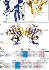
IPSEΔNLS adopts a βγ-crystallin fold. A, zoomed view of IPSEΔNLS (blue, right) and human eye lens γ-B-crystallin (dark yellow, left). Shown are the residues of the tyrosine corner of γ-B-crystallin (Tyr-151 and Arg-147) and the orthogonal stacked Tyr-72 and -74 as well as the disulfide bridges of IPSEΔNLS (sticks) and the flexible loop (amino acids (aa) 62–69) (green). B, structure superposition of two IPSEΔNLS monomers (blue) with dimeric Spherulin 3A (orange). Structures align with a backbone r.m.s.d. of 1.8 Å. C, structure-based multiple sequence alignment of crystallin superfamily members. Above the sequences: secondary structure elements of IPSEΔNLS (except of disulfide bridges). Below the sequences: disulfide bridges are shown as red numbers; the flexible loop (amino acids 62–69) unique to IPSE/α-1 is depicted as a green bar; the two (F/Y)XXXX(F/Y)G signature sequences and the conserved serine residues characteristic of the crystallin fold are indicated by asterisks.
FIGURE 4.
FIGURE 4.
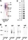
IPSE/α-1 is an immunoglobulin-binding protein with highest affinity to IgE. A, IPSE/α-1 binds to immunoglobulins and transferrin (Tf) in human serum. Western blots of human serum were incubated with IPSE/α-1 followed by labeled streptavidin (SAV-AP) or labeled α-human IgG. Proteins were stained with AuroDye and india ink. B, IPSE/α-1 binds to various immunoglobulin isotypes. Strips with blotted human polyclonal IgE, IgG, IgA, and IgM were incubated with the respective anti-isotype antibodies or IPSE/α-1 followed by SAV-AP. Controls: secondary antibody (C1), SAV-AP (C2). C, dotted IgG (G) and IgE (E) were incubated with labeled IPSE/α-1 followed by SAV-AP.
FIGURE 5.
FIGURE 5.
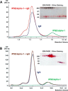
Size exclusion chromatography. Complexes of IPSE/α-1 and IgE (A) and complexes of IPSE/α-1 and IgG (ratio 1:1), respectively (B), were applied to a Superdex 75 column (red curve). For control, the same amount IPSE/α-1 alone (green curve) and IgE or IgG alone (blue curve) were run over the column. Fractions of the IPSE/α-1·IgE or IPSE/α-1·IgG complexes were analyzed by SDS-PAGE and silver staining. High molecular weight fractions contain IPSE/α-1 and IgE but not IgG, suggesting that IPSE/α-1 binds with higher affinity to IgE than to IgG.
FIGURE 6.
FIGURE 6.
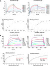
Surface plasmon resonance assays. Binding of IPSE/α-1 dimer (A) and IPSEΔNLS monomer (B) to IgE and IgG immobilized on a CM5 chip was analyzed by real time measurement. Curves were obtained after subtraction of the unspecific binding to the reference cell. Bound IPSE (RU) was estimated at the end of the association phase. Binding isotherms of the IPSE/α-1 dimer (A) showed a sigmoidal curve characteristic for multivalent positive cooperative binding. Thus, Langmuir (1:1) calculation of affinity constants was not applicable. For the IPSEΔNLS monomer (B) dissociation constants of 1.7 and 7.4 μm for IgE and IgG, respectively, were measured.
FIGURE 7.
FIGURE 7.
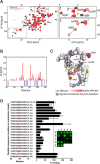
Mapping the binding surface of IPSEΔNLS with IgE. A, 1H,15N NMR correlation spectra of IPSEΔNLS (black) and the IPSEΔNLS·IgE complex (molecular mass ∼200 kDa, red). The presence of NMR signals corresponding to free IPSEΔNLS at a molar ratio of ∼1:1 (IPSEΔNLS/IgE) suggests that IgE has only one IPSEΔNLS-binding site. (Note, the NMR signals of even a very small fraction of free IPSEΔNLS (10 kDa) are much stronger than those of the 200-kDa complex.) B, NMR chemical shift perturbation (CSP) versus residue number. Blue negative bars indicate amide signals that are broadened beyond detection. C, mapping of the IgE binding interface onto the structure of IPSEΔNLS. Nitrogen atoms of the backbone amide groups are represented as spheres and colored red according to CSP or blue if line broadening is observed in the NMR titration experiment. D, peptide scan. Binding of labeled IgE was to immobilized 15-mer IPSE-derived peptides. Binding (means ± S.E.) of IgE-DY781 to 15-mer peptides is expressed in relation to the IPSE-derived peptide 62–76 (inset, spots D1 and D2; mean fluorescence = 100%). Residues exchanged to Ala or Glu in the respective peptides are highlighted in red. Inset, representative example of a peptide library read-out.
FIGURE 8.
FIGURE 8.
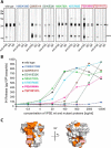
Mutational analysis of immunoglobulin binding and functional activity of IPSE/α-1. A, binding of monoclonal anti-IPSE antibody, human IgG, or human IgE to IPSE/α-1 wild-type and mutants, respectively, assessed by Western blots. Mutants in red boxes were analyzed by SPR for binding affinity with IgE. B, IL-4 release from human basophils induced by different concentrations of IPSE/α-1 wild-type and mutants, respectively. Shown is one representative result of three independent experiments. C, surface representation of IPSEΔNLS. Binding interface was derived from the NMR titration and the peptide scan (orange).
FIGURE 9.
FIGURE 9.
One-dimensional 1H NMR (A) and 1H,15N HSQC spectra (B) spectra of IPSEΔNLS and IPSEΔNLS mutants. A, spectral regions of amide and aromatic and methyl protons for the mutants Q28R/E41K, K67E/R68E, F62A/N64A, I108E/K109E, and D31K/E32K are shown. The good dispersion of amide proton signals and the presence of methyl proton signals below 0 ppm observed for all variants except I108E/K109E are indicative of the presence of tertiary folds. Signals arising from low molecular weight contaminations are marked with an asterisk. Spectra were recorded at 600 MHz 1H Larmor frequency. B, only signals of amides in close spatial proximity to the mutations are shifted in the spectra of D31K/E32K and K67E/R68E mutants. Spectra were recorded at 600 MHz 1H Larmor frequency.
FIGURE 10.
FIGURE 10.
Mode of interaction of IPSE/α-1 with immunoglobulins. A, binding of IPSE/α-1 to deglycosylated IgG Fc. Blotted mock-treated IgG Fc (Control) and IgG Fc deglycosylated with PNGase F were incubated with α-human IgG Fc or A. aurantia agglutinin or labeled IPSE/α-1. B, IPSE/α-1 binds to Fab and Fc fragments. Blotted Fab and Fc fragments of human IgG were detected by α-human IgG (Fc), by α-κ light chain (anti-Ig kappa), by A. aurantia agglutinin or by labeled IPSE/α-1. C1 = buffer control and C2 = SAV-AP. C, sandwich blots. Human IgE dotted at different amounts (10 to 0.1 pmol) was incubated with buffer (negative control) or α-IgE (positive control) or IPSE/α-1, followed by biotinylated IgE. α-IgE gives a clear signal with a maximum at 0.3 pmol/dot, whereas IPSE/α-1 does not exceed background level. D, double immunodiffusion assay (Ouchterlony). Binding partners were applied at various ratios. One partner was pipetted into the center well, the other one clockwise beginning at 12 o'clock in order of dilution in the surrounding wells: 1:1, 1:2, 1:4, 1.8, 1:16, and 1:32. The following starting concentrations were used: 0.5 mg/ml IgE and 1 mg/ml anti-IgE; 0.5 mg/ml IgG and 0.5 mg/ml anti-IgG; 0.5 mg/ml IgG and 0.5 mg/ml protein L (a B cell superantigen from P. magnus); 0.25 mg/ml IgE and 0.05 mg/ml IPSE/α-1 (corresponds to a molar ratio of ∼1:1); 0.25 mg/ml IgG and 0.05 mg/ml IPSE/α-1; and 0.05 mg/ml IPSE/α-1 and rabbit anti-IPSE antiserum (1:2). Precipitation arcs indicate complex formation.
FIGURE 11.
FIGURE 11.
Model of the interaction of IPSE/α-1 with IgE. A putative novel mechanism of basophil activation is shown. A, surface representation of IgE Fc (PDB accession code 2Y7Q, left) and IPSEΔNLS (right) colored according to electrostatic surface potential at ±2 kB T/e for positive (blue) or negative (red) charge potential using the program APBS (48). B, proposed interaction between IPSEΔNLS and IgE (represented by the crystal structure of IgE Fc/FcϵRI, PDB accession code 2WQR). The positively charged binding interface of IPSEΔNLS mapped by NMR and mutational analysis presumably interacts with a negatively charged surface on the Cϵ2 domain of IgE, while the FcϵRI binds to the neighboring Cϵ3 domain of the IgE molecule. C, schematic model of IgE receptor activation on the surface of basophils. IPSE/α-1 may induce a structural rearrangement in the IgE molecule. This triggers the activation of the receptor signaling cascade.

Similar articles

Cited by

References

    1. Schramm G., Haas H. (2010) Th2 immune response against Schistosoma mansoni infection. Microbes Infect. 12, 881–888 - PubMed
    1. Seder R. A., Paul W. E., Davis M. M., Fazekas de St Groth B. (1992) The presence of interleukin 4 during in vitro priming determines the lymphokine-producing potential of CD4+ T cells from T cell receptor transgenic mice. J. Exp. Med. 176, 1091–1098 - PMC - PubMed
    1. Brunet L. R., Finkelman F. D., Cheever A. W., Kopf M. A., Pearce E. J. (1997) IL-4 protects against TNF-α 32 -mediated cachexia and death during acute schistosomiasis. J. Immunol. 159, 777–785 - PubMed
    1. Herbert D. R., Orekov T., Perkins C., Rothenberg M. E., Finkelman F. D. (2008) IL-4Rα expression by bone marrow-derived cells is necessary and sufficient for host protection against acute schistosomiasis. J. Immunol. 180, 4948–4955 - PMC - PubMed
    1. Sullivan B. M., Liang H. E., Bando J. K., Wu D., Cheng L. E., McKerrow J. K., Allen C. D., Locksley R. M. (2011) Genetic analysis of basophil function in vivo. Nat. Immunol. 12, 527–535 - PMC - PubMed

Publication types

MeSH terms