. 2015 Oct;1847(10):1245-53.
doi: 10.1016/j.bbabio.2015.07.002. Epub 2015 Jul 9.

Calcium-induced conformational changes in the regulatory domain of the human mitochondrial ATP-Mg/Pi carrier

Affiliations

Calcium-induced conformational changes in the regulatory domain of the human mitochondrial ATP-Mg/Pi carrier

Steven P D Harborne et al. Biochim Biophys Acta. 2015 Oct.

Abstract

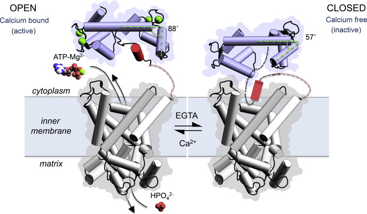
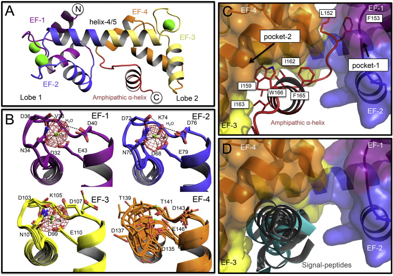
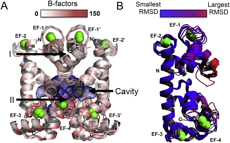
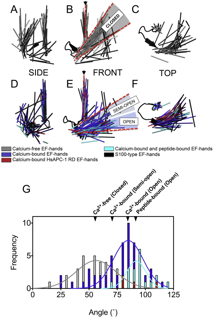
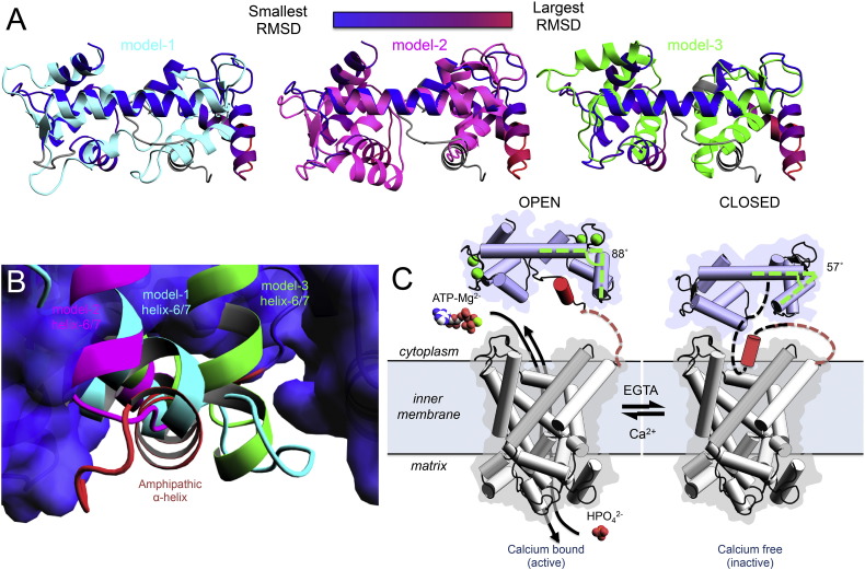
The mitochondrial ATP-Mg/Pi carrier imports adenine nucleotides from the cytosol into the mitochondrial matrix and exports phosphate. The carrier is regulated by the concentration of cytosolic calcium, altering the size of the adenine nucleotide pool in the mitochondrial matrix in response to energetic demands. The protein consists of three domains; (i) the N-terminal regulatory domain, which is formed of two pairs of fused calcium-binding EF-hands, (ii) the C-terminal mitochondrial carrier domain, which is involved in transport, and (iii) a linker region with an amphipathic α-helix of unknown function. The mechanism by which calcium binding to the regulatory domain modulates substrate transport in the carrier domain has not been resolved. Here, we present two new crystal structures of the regulatory domain of the human isoform 1. Careful analysis by SEC confirmed that although the regulatory domain crystallised as dimers, full-length ATP-Mg/Pi carrier is monomeric. Therefore, the ATP-Mg/Pi carrier must have a different mechanism of calcium regulation than the architecturally related aspartate/glutamate carrier, which is dimeric. The structure showed that an amphipathic α-helix is bound to the regulatory domain in a hydrophobic cleft of EF-hand 3/4. Detailed bioinformatics analyses of different EF-hand states indicate that upon release of calcium, EF-hands close, meaning that the regulatory domain would release the amphipathic α-helix. We propose a mechanism for ATP-Mg/Pi carriers in which the amphipathic α-helix becomes mobile upon release of calcium and could block the transport of substrates across the mitochondrial inner membrane.

Keywords: Adenine nucleotide translocase; Calcium regulation mechanism; EF-hand conformational change; Regulation of adenine nucleotides; SCaMC.


Figures

None
Graphical abstract
Fig. 1
Fig. 1
Features of the HsAPC-1 RD structure. A) A single chain of the HsAPC-1 regulatory domain (RD) structure. B) Detailed views of the EF-hands where each observation in the seven chains is superposed. Electron density from an anomalous difference Fourier map, calculated from the P212121 space-group, is displayed as a mesh in red (σ-level of 5). C) View of hydrophobic pockets 1 and 2 that bind the loop preceding the amphipathic α-helix and the amphipathic α-helix itself, respectively, or D) the position of calmodulin recognition sequence motifs (black for calmodulin peptides or cyan for others) when EF-hands of other proteins are superposed on lobe 2 of the HsAPC-1 RD structure. In panels A and B a cartoon representation of the HsAPC-1 RD molecule is shown, but in panels C and D a surface representation is used. In all panels EF-hands 1 to 4 are coloured in purple, blue, yellow and orange respectively. The amphipathic α-helix is represented in red. Calcium ions are represented as lime green spheres, and red spheres represent water molecules. In all cases, where side chains are shown, they are represented as sticks, and nitrogen and oxygen atoms are coloured according to convention.
Fig. 2
Fig. 2
Crystal packing of HsAPC-1 RD chains, and comparison to previous HsAPC-1 RD structure. A) Seven unique dimer combinations of HsAPC-1 RD in total were superposed upon one another. Each dimer is represented as a cartoon and coloured according to B-factor values as indicated in the key. The two main points of contact between each chain of the dimer are labelled I and II respectively. B) The seven observed chains of the HsAPC-1 RD were superposed on the previously published one (PDB ID: 4N5X). The structures in panel B are coloured by the RMSD difference from 4N5X as in the key. Calcium ions are represented as lime green spheres.
Fig. 3
Fig. 3
Full-length HsAPC-1 is monomeric. A) SEC trace for full-length HsAPC-1 and protein quantification by SDS-PAGE gel (inset). B) Contributions of protein, detergent and lipid to the total mass. C) SEC trace for crudely isolated HsAPC-1 RD. Peak 1 was found to contain a contaminant, and identified as the 56.1 kDa E2 component of the L. lactis pyruvate dehydrogenase complex (Supplementary Fig. 1). In panels A and C the elution volume of molecular weight standards are indicated and annotated with the mass of the standard. D) Protein from each of the peaks 2 and 3 run on an SDS-PAGE gel in the presence or absence of DTT.
Fig. 4
Fig. 4
Calcium-induced conformational changes in EF-hands. Views of 129 EF-hands superposed on EF-hand 1 of calcium-bound calmodulin, in the calcium-free (A–C) and calcium-bound (D–F) states. Entering and exiting α-helices are represented as a line though the α-helix centre. G) A histogram of the angles between the entering and exiting α-helices of conventional (not S100-like) EF-hands.
Fig. 5
Fig. 5
A model for the calcium regulation of HsAPC-1 RD. A) Three models for calcium-free HsAPC-1 RD based on centrin (model-1), calmodulin (model-2) and calcium-dependent protein kinase (model-3) were superposed onto the calcium-bound HsAPC-1 RD structure and coloured cyan, magenta and green respectively. The calcium-bound HsAPC-1 structure is coloured by RMSD difference from the calcium-free models as in the key. B) A close up view of hydrophobic pocket 2 into which the amphipathic α-helix (red) is bound. The calcium-bound HsAPC-1 RD structure is shown as a surface representation and coloured blue, whereas the superposed calcium-free models are represented as cartoon (coloured as in panel A). C) The proposed regulatory mechanism between calcium-bound state of HsAPC-1 where the carrier is active and the calcium-free state of HsAPC-1 where the carrier is inactive. The calcium-free model presented in panel C is based on model 2. α-helices are represented as cylinders in panel C. Calcium ions are presented as lime green spheres.

Similar articles

Cited by

References

    1. Palmieri F. The mitochondrial transporter family SLC25: identification, properties and physiopathology. Mol. Asp. Med. 2013;34:465–484. - PubMed
    1. Austin J., Aprille J.R. Carboxyatractyloside-insensitive influx and efflux of adenine nucleotides in rat liver mitochondria. J. Biol. Chem. 1984;259:154–160. - PubMed
    1. Fiermonte G., De Leonardis F., Todisco S., Palmieri L., Lasorsa F.M., Palmieri F. Identification of the mitochondrial ATP-Mg/Pi transporter. Bacterial expression, reconstitution, functional characterization, and tissue distribution. J. Biol. Chem. 2004;279:30722–30730. - PubMed
    1. Cavero S., Traba J., del Arco A., Satrustegui J. The calcium-dependent ATP-Mg/Pi mitochondrial carrier is a target of glucose-induced calcium signalling in Saccharomyces cerevisiae. Biochem. J. 2005;392:537–544. - PMC - PubMed
    1. Klingenberg M. The ADP and ATP transport in mitochondria and its carrier. Biochim. Biophys. Acta Biomembr. 2008;1778:1978–2021. - PubMed

LinkOut - more resources