. 2015 Jul 23;22(7):946-56.
doi: 10.1016/j.chembiol.2015.06.012. Epub 2015 Jul 9.

A 45-Amino-Acid Scaffold Mined from the PDB for High-Affinity Ligand Engineering

Affiliations

A 45-Amino-Acid Scaffold Mined from the PDB for High-Affinity Ligand Engineering

Max A Kruziki et al. Chem Biol. .

Abstract

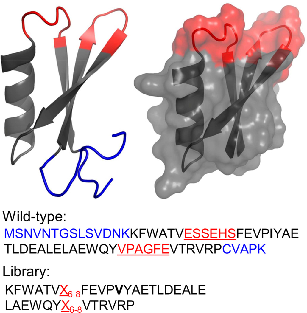
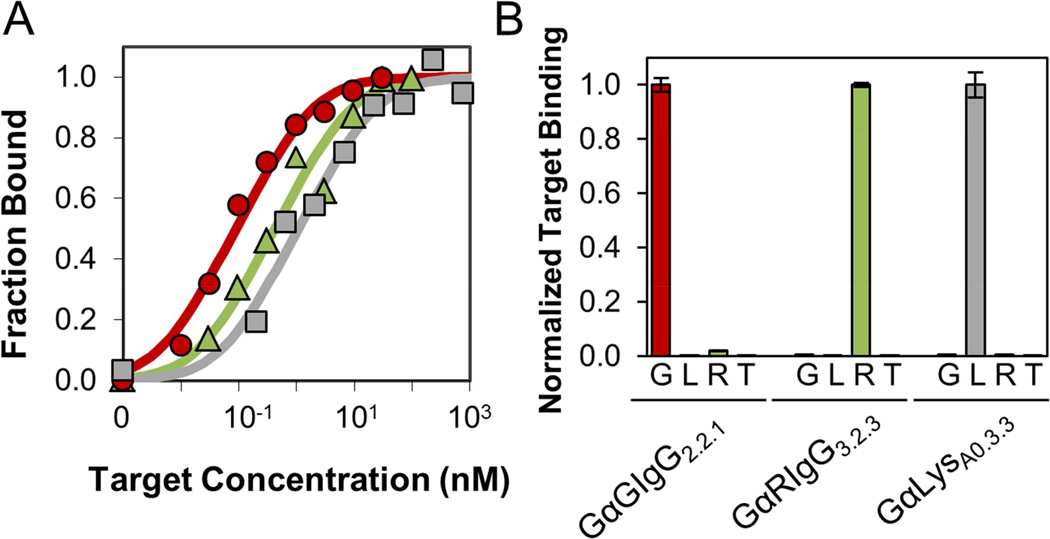
Small protein ligands can provide superior physiological distribution compared with antibodies, and improved stability, production, and specific conjugation. Systematic evaluation of the PDB identified a scaffold to push the limits of small size and robust evolution of stable, high-affinity ligands: 45-residue T7 phage gene 2 protein (Gp2) contains an α helix opposite a β sheet with two adjacent loops amenable to mutation. De novo ligand discovery from 10(8) mutants and directed evolution toward four targets yielded target-specific binders with affinities as strong as 200 ± 100 pM, Tms from 65 °C ± 3 °C to 80°C ± 1 °C, and retained activity after thermal denaturation. For cancer targeting, a Gp2 domain for epidermal growth factor receptor was evolved with 18 ± 8 nM affinity, receptor-specific binding, and high thermal stability with refolding. The efficiency of evolving new binding function and the size, affinity, specificity, and stability of evolved domains render Gp2 a uniquely effective ligand scaffold.


Figures

Figure 1
Figure 1. Summary of information for top potential scaffolds
(A) Structure and proposed paratope surface (red) of top five scoring scaffolds. Images created in Pymol. (B) PDB ID, amino acid length, % β strand, and % α helix, are all from the Protein Data Bank. Loop residues are identified as residues between secondary structural elements. Loop accessible surface area (ASA) is calculated using GetArea after mutating loop residues to serines within PyMol. ΔΔGf;mut was calculated for random loop mutants using Eris. Fibronectin (l ttg) and Affibody (2b88) are included for comparison. Also see Table S1 for commonly used scaffolds.
Figure 2
Figure 2. Solution structure of Gp2
Diversified amino acids are highlighted in red and underlined in sequence. The N- and C- terminal tails removed to create the Gp2 scaffold are highlighted in blue. An I17V (boldface in sequence) mutation was added based on prevalence in homologous protein sequences. Images created in Macpymol (PDB ID: 2wnm).
Figure 3
Figure 3. Binding characterization
(A) Yeast displaying GαGIgG2.2.1 (red circles), GαRIgG3.2.3 (green triangles), and GαLysA0.3.3 (gray squares) were incubated with the indicated concentrations of biotinylated target. Binding was detected by streptavidin-fluorophore via flow cytometry. Titration indicates equilibrium dissociation constants of 0.2 ±0.1 nM, 2.3 ±1.4 nM, and 0.9 ±0.7 nM. (B) Yeast were incubated with 1 µM of goat IgG (G), lysozyme (L), rabbit IgG (R), or transferrin (T). Binding was detected by streptavidin-fluorophore via flow cytometry. Data are normalized to signal of intended target for each yeast strain. Error bars represent ±SD of n = 3 samples. See Figure S3 for full collection of affinity titration curves.
Figure 4
Figure 4. Soluble Gp2 characterization
(A,B) Purified Gp2 clones (blue: wild-type, red: GαGIgG2.2.1, green: GαRIgG3.2.3) were analyzed by circular dichroism spectroscopy. (A) Molar ellipticity (θ) was measured at indicated wavelengths before (solid lines) and after (dashed lines) thermal denaturation. (B) Molar ellipticity was monitored at 218 nm upon heating from 25 °C to 98 °C at 1 °C per minute. (C) Biotinylated IgG (2 nM goat or 4 nM rabbit as appropriate) was incubated with the indicated concentration of purified Gp2 and used to label yeast displaying the corresponding Gp2 clone. IgG binding was measured by streptavidin-fluorophore via flow cytometry. (D) Purified GαGIgG2.2.1 was subjected to various treatments prior to use in the competitive binding assay (as in (C)). Unblocked is a control without Gp2 competition. Pre-HPLC uses competition by GαGIgG2.2.1 that was purified on a metal affinity column. Post-HPLC uses competition by GαGIgG2.2.1 purified by metal affinity chromatography and reverse phase high performance liquid chromatography and lyophilization. Post-heat uses competition by GαGIgG2.2.1 purified as in Post-HPLC along with heating to 98 °C and cooling back to 22 °C. Error bars are ±SD on n = 2 samples. See Figure S2 for GαLysA0.3.3.
Figure 5
Figure 5. GαEGFR2.2.3 affinity and stability
(A) Population of yeast displaying Gp2 incubated with 50 nM EGFR and anti-c-myc antibody. Secondary fluorophores detect binding of EGFR and antibody. Spread of double positive cells suggests moderate diversity of EGFR binding Gp2 molecules. GαEGFR2.2.3 was collected from top 1% of double positives. (B) A431 cells were incubated with indicated amount of purified Gp2. Binding was detected by anti-His6 fluorophore. Titration indicates an equilibrium dissociation constant of 18 ±8 nM. (C) Molar ellipticity was monitored at 218 nm upon heating from 25 °C to 98 °C at 1 °C per minute. Molar ellipticity before (solid) and after (dashed) thermal denaturation is inset. (D) Fluorescence microscopy of adhered cancer cell lines, incubated with 100 nM Gp2 protein and detected by anti-His6-fluorescein. A431 cells labeled with GαEGFR2.2.3 (left column) show localization to cell surface. A431 cells labeled with Gp2 WT (middle column) and MCF7 cells labeled with GαEGFR2.2.3 (lower column) show very little detectable binding. Scale bar represents 200 µm. (E) A431 cells preincubated with PBS or 1 µM GαEGFR2.2.3 and labeled with 200 nM biotinylated GαEGFR2.2.3. Binding detected with streptavidin fluorophore. (F) Cancer cell lines expressing varying levels of EGFR: MCF7 with 2 × 10⁴ receptors (gray line) (Reilly et al., 2000), MDA-MB-231 with 1 × 10⁵ receptors (red dot) (Reilly et al., 2000), DU145 with 2 × 10⁵ receptors (black dash) (Malmberg et al., 2011), and A431with 3 × 10⁶ (green dash) (Spangler et al., 2010) were incubated with 1 µM GαEGFR2.2.3 and detected with anti-His6-fluorescein by flow cytometry.
Figure 6
Figure 6. Deep sequencing comparison of naïve and binding populations
All sequences were grouped into families and damped. (A) Diversified loop positions (CDR`) are shown at the top, with 9a and 9b, or 36a and 36b, representing loop length diversity positions. Positions with an absolute change of 5% or greater are labeled. All labeled positions have p < 0.001 (n ≥ 96, number of damped sequences). (B) Relative change in amino acid frequency by framework position from the naïve library to combined mature populations. Certain mutations are more common than others due to error prone PCR limitations. Positions where wild type was conserved (#) or mutated (*) significantly more often (p < 0.001 for 3% deviation, n ≥ 421) than the mean mutation rate are denoted. (C) Frequency of Gp2 loop length in amino acids. The naïve library was designed with equal loop length frequencies, but DNA bias during construction lead to overrepresentation of shorter loops. Mature combines high throughput sequencing from the four binding populations. All have p < 0.001. Error bars are ±SD for n = 4.9 × 10⁴ for naïve and n ≥ 477 for mature (number of damped sequences). See also Figure S1.

References

    1. Ackerman SE, Currier NV, Bergen JM, Cochran JR. Cystine-knot peptides: emerging tools for cancer imaging and therapy. Expert Rev. Proteomics. 2014;11:561–572. - PubMed
    1. Banta S, Dooley K, Shur O. Replacing antibodies: engineering new binding proteins. Annu. Rev. Biomed. Eng. 2013;15:93–113. - PubMed
    1. Baum RP, Prasad V, Müller D, Schuchardt C, Orlova A, Wennborg A, Tolmachev V, Feldwisch J. Molecular imaging of HER2-expressing malignant tumors in breast cancer patients using synthetic 111In- or 68Ga-labeled affibody molecules. J. Nucl. Med. 2010;51:892–897. - PubMed
    1. Binz HK, Amstutz P, Plückthun A. Engineering novel binding proteins from nonimmunoglobulin domains. Nat. Biotechnol. 2005;23:1257–1268. - PubMed
    1. Bloom JD, Labthavikul ST, Otey CR, Arnold FH. Protein stability promotes evolvability. Proc. Natl. Acad. Sci. 2006;103:5869–5874. - PMC - PubMed

Publication types