

CD4(+) T cells epigenetically modified by oxidative stress cause lupus-like autoimmunity in mice
- PMID: 26165613
- PMCID: PMC4529773
- DOI: 10.1016/j.jaut.2015.06.004
CD4(+) T cells epigenetically modified by oxidative stress cause lupus-like autoimmunity in mice
Abstract
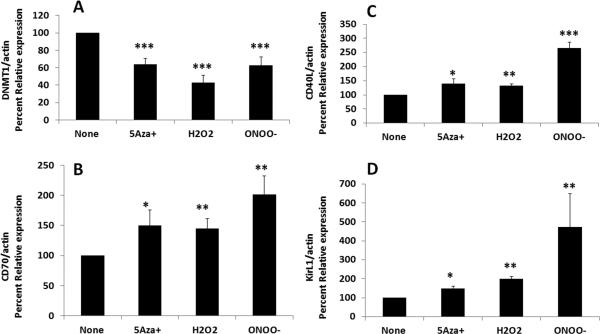
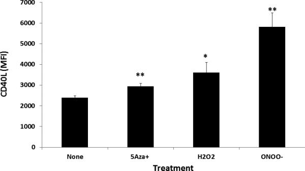
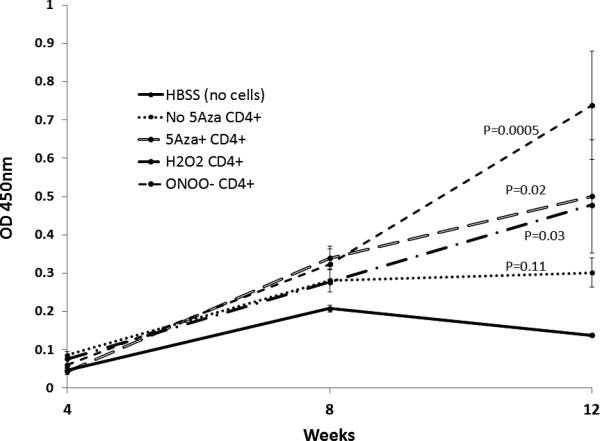
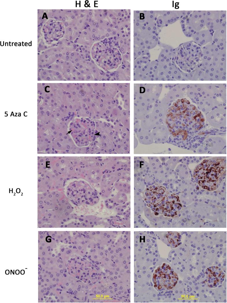
Lupus develops when genetically predisposed people encounter environmental agents such as UV light, silica, infections and cigarette smoke that cause oxidative stress, but how oxidative damage modifies the immune system to cause lupus flares is unknown. We previously showed that oxidizing agents decreased ERK pathway signaling in human T cells, decreased DNA methyltransferase 1 and caused demethylation and overexpression of genes similar to those from patients with active lupus. The current study tested whether oxidant-treated T cells can induce lupus in mice. We adoptively transferred CD4(+) T cells treated in vitro with oxidants hydrogen peroxide or nitric oxide or the demethylating agent 5-azacytidine into syngeneic mice and studied the development and severity of lupus in the recipients. Disease severity was assessed by measuring anti-dsDNA antibodies, proteinuria, hematuria and by histopathology of kidney tissues. The effect of the oxidants on expression of CD40L, CD70, KirL1 and DNMT1 genes and CD40L protein in the treated CD4(+) T cells was assessed by Q-RT-PCR and flow cytometry. H2O2 and ONOO(-) decreased Dnmt1 expression in CD4(+) T cells and caused the upregulation of genes known to be suppressed by DNA methylation in patients with lupus and animal models of SLE. Adoptive transfer of oxidant-treated CD4(+) T cells into syngeneic recipients resulted in the induction of anti-dsDNA antibody and glomerulonephritis. The results show that oxidative stress may contribute to lupus disease by inhibiting ERK pathway signaling in T cells leading to DNA demethylation, upregulation of immune genes and autoreactivity.
Keywords: CD40L; CD70; KirL1; Systemic lupus erythematosus (SLE).
Copyright © 2015 Elsevier Ltd. All rights reserved.
Figures
Similar articles
-
Oxidative stress, T cell DNA methylation, and lupus.Arthritis Rheumatol. 2014 Jun;66(6):1574-82. doi: 10.1002/art.38427. Arthritis Rheumatol. 2014. PMID: 24577881 Free PMC article.
-
Overexpression of the growth arrest and DNA damage-induced 45alpha gene contributes to autoimmunity by promoting DNA demethylation in lupus T cells.Arthritis Rheum. 2010 May;62(5):1438-47. doi: 10.1002/art.27363. Arthritis Rheum. 2010. PMID: 20131288
-
The interaction between environmental triggers and epigenetics in autoimmunity.Clin Immunol. 2018 Jul;192:1-5. doi: 10.1016/j.clim.2018.04.005. Epub 2018 Apr 9. Clin Immunol. 2018. PMID: 29649575 Free PMC article. Review.
-
Treating activated CD4+ T cells with either of two distinct DNA methyltransferase inhibitors, 5-azacytidine or procainamide, is sufficient to cause a lupus-like disease in syngeneic mice.J Clin Invest. 1993 Jul;92(1):38-53. doi: 10.1172/JCI116576. J Clin Invest. 1993. PMID: 7686923 Free PMC article.
-
Epigenetic regulation and the pathogenesis of systemic lupus erythematosus.Transl Res. 2009 Jan;153(1):4-10. doi: 10.1016/j.trsl.2008.10.007. Epub 2008 Nov 14. Transl Res. 2009. PMID: 19100952 Review.
Cited by
-
Chronic exposure to water pollutant trichloroethylene increased epigenetic drift in CD4(+) T cells.Epigenomics. 2016 May;8(5):633-49. doi: 10.2217/epi-2015-0018. Epub 2016 Apr 19. Epigenomics. 2016. PMID: 27092578 Free PMC article.
-
Epigenetically Altered T Cells Contribute to Lupus Flares.Cells. 2019 Feb 5;8(2):127. doi: 10.3390/cells8020127. Cells. 2019. PMID: 30764520 Free PMC article. Review.
-
Continuous Developmental and Early Life Trichloroethylene Exposure Promoted DNA Methylation Alterations in Polycomb Protein Binding Sites in Effector/Memory CD4+ T Cells.Front Immunol. 2019 Aug 28;10:2016. doi: 10.3389/fimmu.2019.02016. eCollection 2019. Front Immunol. 2019. PMID: 31555266 Free PMC article.
-
The relationship of polluted air and drinking water sources with the prevalence of systemic lupus erythematosus: a provincial population-based study.Sci Rep. 2021 Sep 20;11(1):18591. doi: 10.1038/s41598-021-98111-8. Sci Rep. 2021. PMID: 34545152 Free PMC article.
-
Autoimmunity in 2015.Clin Rev Allergy Immunol. 2016 Aug;51(1):110-9. doi: 10.1007/s12016-016-8576-1. Clin Rev Allergy Immunol. 2016. PMID: 27422713 Review.
References
-
- Quddus J, Johnson KJ, Gavalchin J, Amento EP, Chrisp CE, Yung RL, et al. Treating activated CD4+ T cells with either of two distinct DNA methyltransferase inhibitors, 5-azacytidine or procainamide, is sufficient to cause a lupus-like disease in syngeneic mice. The Journal of clinical investigation. 1993;92:38–53. - PMC - PubMed
-
- Gorelik G, Fang JY, Wu A, Sawalha AH, Richardson B. Impaired T cell protein kinase C delta activation decreases ERK pathway signaling in idiopathic and hydralazine-induced lupus. Journal of immunology. 2007;179:5553–63. - PubMed
Publication types
MeSH terms
Substances
Grants and funding
LinkOut - more resources
Full Text Sources
Other Literature Sources
Research Materials
Miscellaneous
