. 2015 Jul 16;162(2):287-299.
doi: 10.1016/j.cell.2015.06.014. Epub 2015 Jul 9.

GM130 Regulates Golgi-Derived Spindle Assembly by Activating TPX2 and Capturing Microtubules

Affiliations

GM130 Regulates Golgi-Derived Spindle Assembly by Activating TPX2 and Capturing Microtubules

Jen-Hsuan Wei et al. Cell. .

Abstract

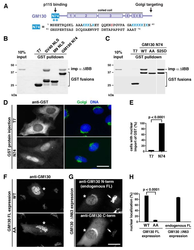
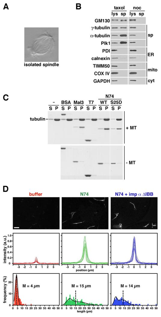
Spindle assembly requires the coordinated action of multiple cellular structures to nucleate and organize microtubules in a precise spatiotemporal manner. Among them, the contributions of centrosomes, chromosomes, and microtubules have been well studied, yet the involvement of membrane-bound organelles remains largely elusive. Here, we provide mechanistic evidence for a membrane-based, Golgi-derived microtubule assembly pathway in mitosis. Upon mitotic entry, the Golgi matrix protein GM130 interacts with importin α via a classical nuclear localization signal that recruits importin α to the Golgi membranes. Sequestration of importin α by GM130 liberates the spindle assembly factor TPX2, which activates Aurora-A kinase and stimulates local microtubule nucleation. Upon filament assembly, nascent microtubules are further captured by GM130, thus linking Golgi membranes to the spindle. Our results reveal an active role for the Golgi in regulating spindle formation to ensure faithful organelle inheritance.


Figures

Figure 1
Figure 1
Acute Perturbation of GM130 in Mitosis Impairs Spindle Assembly and Cell Division (A) Microinjection of antibodies against the N-terminal domain of GM130 (residues 1-74, N74) interferes with bipolar division. NRK cells in pro/prometaphase were injected with 9 mg/ml affinity-purified anti-N74 or control IgG together with fluorescent dextran as an injection marker. Cell division was then assessed by fluorescence microscopy. (B) Anti-N74 injection decreased bipolar division to 38% compared to control IgG. 373 (ctl IgG) and 311 (anti-N74) cells from 4 experiments were analyzed, p<0.0001. (C) Microinjection of N74 protein interferes with bipolar division. Recombinant T7-GST, N74-GST or GST-SV40 NLS was injected as in (A). Mitotic progression was then monitored by phase-contrast time-lapse microscopy. T7 and SV40-injected cells progressed through mitosis and divided normally. N74-injected cells frequently stalled in mitosis and died after a prolonged arrest. (D) N74 injection decreased bipolar division to 44%, while T7 and SV40 had little effect. 83 (T7), 81 (N74), and 138 (SV40) cells from 3-4 experiments were analyzed, p<0.005. (E) Microinjection of N74 induces spindle assembly defects. Injected cells were stained for DNA and α-tubulin (MT). N74-injected cells showed various defects in chromosome alignment and spindle assembly. Notably, category II phenocopied RanQ69L-injection that induced ectopic asters. (F) Percentage of mitotic phenotypes in injected cells. 75 (T7), 201 (N74), and 190 (SV40) cells from 3-6 experiments were analyzed. Bars, 25 μm. Error bars represent SEM. See also Figure S1.
Figure 2
Figure 2
GM130 Harbors a bona fide Classical NLS (A) Domain structure. GM130 is comprised of six coiled-coil regions in the middle, a Golgi-targeting domain at the C-terminus and a p115-interacting motif at the N-terminus (N74). N74 contains two K/R-rich clusters (blue) and a Cdk1 phosphorylation site S25 (red). (B) GM130 N74 directly interacts with importin α in vitro. Upon incubation with the NLS-interacting domain of importin α (ΔIBB), GST fusion proteins were pulled down and analyzed by SDS-PAGE and Coomassie blue staining. N74 and the classical SV40 NLS bound importin α, but not T7 or the non-classical M9 NLS. (C) Pulldowns were performed as in (B) with the N74 NLS mutant AA (K/R clusters changed to alanine) or the Cdk1 phospho-mimicking mutant S25D. Importin α binding required the K/R residues and was not affected by S25 phospho-mimicking mutation. The blank space indicates removal of irrelevant lanes. (D) N74 is recognized and translocated into the nucleus by the endogenous nuclear import machinery in cells. NRK cells were microinjected into the cytoplasm with T7-GST or N74-GST and then stained for DNA, GST, and the Golgi (with an antibody against the C-terminal domain of GM130). (E) N74-GST was imported into the nucleus in 99.7% of the injected cells. 379 (T7) and 358 (N74) cells from 3 experiments were analyzed, p<0.0001. (F) Over-expressed full-length GM130 targets to the Golgi and enters the nucleus. 93% of GM130 FL WT- and 6% of GM130 FL AA-expressing cells (marked by asterisks) showed nuclear localization of GM130. (G) Endogenous GM130 translocates into the nucleus once mobilized. Upon expression of a Golgi-targeting, N-terminal truncation mutant of GM130 (ΔN63), endogenous GM130 was displaced from the Golgi and entered the nucleus in 87% of ΔN63-expressing cells, as detected with an antibody against the N-terminal domain. Expressing and non-expressing cells are marked by asterisks and arrows, respectively. (H) Quantitation of (F) and (G). 170 (FL WT), 167 (FL AA), and 358 (ΔN63) cells from 3-4 experiments were analyzed, p<0.0001. Bars, 25 μm. Error bars represent SEM. See also Figure S2.
Figure 3
Figure 3
Importin α Is Recruited to Mitotic Golgi Membranes by GM130 (A) Importin α is partially membrane-bound. A post-nuclear supernatant (PNS) from unsynchronized HeLa cells was fractionated into cytosol (C) and membranes (M). Equal-volume fractions were analyzed by Western blotting. While importin β was restricted to the cytosol, a portion of importin α bound to membranes. (B) Membrane-bound importin α is elevated in mitosis. Equal protein amounts of membrane fractions from interphase and mitotic HeLa cells were analyzed by Western blotting. In contrast to the unaltered levels of membrane markers, including GM130 and giantin (Golgi), calnexin and PDI (ER), TIMM50 (mitochondria) and membrane-bound γ-tubulin, membrane association of importin α increased in mitosis. (C) Importin α co-sediments with mitotic Golgi membranes. A post-chromosomal supernatant (PCS) from mitotic HeLa cells was centrifuged through a glycerol gradient. Membranes from each fraction were re-isolated and analyzed by Western blotting. Importin α co-migrated with the Golgi markers GM130 and GRASP65 in the top fractions, while ER (calnexin) and mitochondria (TIMM50) markers were found in the bottom. (D) Pre-incubation of the PCS with N74 WT-GST or anti-N74, but not T7-GST, N74 AA-GST or pre-immune serum, selectively removed the pool of Golgi-bound importin α in the top fractions. Input: 2-4% PCS. n=3, **p<0.01. (E) Importin α forms a complex with GM130 specifically in mitosis. Endogenous GM130 from interphase and mitotic HeLa lysates was immunoprecipitated and analyzed by Western blotting. Endogenous importin α co-precipitated with GM130 from mitotic but not interphase lysates. Error bars represent SEM. See also Figure S3.
Figure 4
Figure 4
GM130 Competes Importin α off TPX2 through its High Affinity NLS (A) Mapping of the NLS in mouse TPX2 (NP_082385.3) by protein sequence alignment with frog TPX2 (AAH68637). NLS-containing peptides (residues 270-289 in frog; 302-322 in mouse) are grey-shaded. The key NLS residues (K284, R285 in frog; K315, K316 in mouse) are highlighted in bold. (B) TPX2 binding to importin α requires a functional NLS. T7-GST, GST-TPX2 NLS wildtype (wt), and GST-TPX2 NLS mutant (K315A K316A, mut) were incubated with importin α ΔIBB, pulled down and analyzed by SDS-PAGE and Coomassie blue staining. Importin α only bound the wt NLS of TPX2 but not the NLS mutant. (C) N74 WT and the phospho-mimicking S25D mutant bind importin α with a nine-fold higher affinity than TPX2. The dissociation constants (Kd, represented as mean ± SEM) were measured by ITC. n≥3. (D) GST-pulldown competition assay. N74 WT but not AA competes importin α off the preformed importin α-TPX2 complex. (E) N74 does not directly bind TPX2-NLS. T7-GST and GST-TPX2 NLS were incubated with N74 WT-His or importin α ΔIBB, pulled down and analyzed by SDS-PAGE and Coomassie blue staining. GST-TPX2 NLS bound importin α ΔIBB but not N74-His.
Figure 5
Figure 5
GM130 Induces Aurora-A Activation and Triggers Aster Formation by Liberating TPX2 (A and B) N74 WT, but not N74 AA or SV40 NLS, stimulates aster formation in mitotic extracts. GST fusion proteins immobilized on beads (A) or as soluble proteins (B) were incubated with mitotic HeLa extracts and fluorescent tubulin. Microtubule asters and autofluorescent beads were detected in the same channel by fluorescence microscopy. The inset in (B) shows a magnified representative aster. (C and E) Aster formation is triggered by mitotic Golgi membranes and blocked by antibodies against N74. The activity was also abolished if Cdk1 was inhibited during pre-treatment of the Golgi. (D) N74-stimulated aster assembly was blocked by addition of importin α ΔIBB or full-length importins α/ß. n=3, ****p<0.0001. (F) N74-stimulated aster formation requires TPX2. N74 failed to induce asters in TPX2-immunodepleted (ID) mitotic extracts. n=3, ****p<0.0001. (G) Immunodepletion of TPX2. Mitotic HeLa extracts were incubated for three consecutive rounds with protein A-conjugated beads pre-coupled to control IgG or anti-TPX2. Extracts and beads were analyzed by Western blotting with antibodies against α-tubulin and TPX2. After the third round, TPX2 was depleted from the mitotic extracts. (H) N74 activates Aurora-A, a downstream target of TPX2. Upon incubation with mitotic cytosol and microtubules, N74-WT but not AA induced Aurora-A T288 phosphorylation (left panel). Addition of importins α/β (middle panels) or antibodies against N74 (Ab, right panel) blocked Aurora-A activation. Bars, 20 μm. Error bars represent SEM. See also Figure S4.
Figure 6
Figure 6
GM130 Associates with the Spindle and Directly Binds to Microtubules (A and B) GM130 co-purifies with spindles. Spindles isolated from mitotic HeLa cells were visualized by DIC microscopy (A) or analyzed by Western blotting (B). Whole cell lysates (lys) and isolated spindles (sp) were probed for the indicated marker proteins. Nocodazole (noc) treatment served as a negative control. (C) N74 binds directly to microtubules. Preformed microtubules were incubated with the indicated proteins and then pelleted by centrifugation. Equal volumes of the supernatant (S) and pellet (P) fractions were analyzed by SDS-PAGE and Coomassie blue staining. N74 WT, N74 S25D, and the microtubule binding protein Mal3/EB1 co-pelleted with microtubules, but not T7-GST or BSA. (D) N74 promotes microtubule bundling. Fluorescently-labeled microtubules treated with buffer, N74, or importin α ΔIBB-prebound N74 were visualized by fluorescence microscopy (top). Intensity line-scans from 10 cross-sections of microtubule structures were superimposed (middle) and length distribution was plotted (bottom, 204 structures on average per condition). M represents mean. Bar, 10 μm.
Figure 7
Figure 7
Model of GM130-Mediated, Golgi-Based Microtubule Assembly in Mitosis ❶Cdk1 phosphorylates GM130 upon mitotic entry; ❷ p115 dissociates from GM130; ❸ GM130 sequesters importin α to mitotic Golgi membranes; ❹ TPX2 is released to stimulate microtubule nucleation and growth in the proximity; ❺ GM130 captures and stabilizes nascent microtubules, thereby anchoring mitotic Golgi clusters to the spindle.

Similar articles

Cited by

References

    1. Baschieri F, Confalonieri S, Bertalot G, Di Fiore PP, Dietmaier W, Leist M, Crespo P, Macara IG, Farhan H. Spatial control of Cdc42 signalling by a GM130-RasGRF complex regulates polarity and tumorigenesis. Nat. Commun. 2014;5:4839. - PMC - PubMed
    1. Basto R, Lau J, Vinogradova T, Gardiol A, Woods CG, Khodjakov A, Raff JW. Flies without centrioles. Cell. 2006;125:1375–1386. - PubMed
    1. Bayliss R, Sardon T, Vernos I, Conti E. Structural basis of Aurora-A activation by TPX2 at the mitotic spindle. Mol. Cell. 2003;12:851–862. - PubMed
    1. Bazzi H, Anderson KV. Acentriolar mitosis activates a p53-dependent apoptosis pathway in the mouse embryo. Proc. Natl. Acad. Sci. U. S. A. 2014;111:E1491–E1500. - PMC - PubMed
    1. Carazo-Salas RE, Guarguaglini G, Gruss OJ, Segref A, Karsenti E, Mattaj IW. Generation of GTP-bound Ran by RCC1 is required for chromatin-induced mitotic spindle formation. Nature. 1999;400:178–181. - PubMed

Publication types