

The Stem Cell Marker Bmi-1 Is Sensitive in Identifying Early Lesions of Carcinoma ex Pleomorphic Adenoma
- PMID: 26166073
- PMCID: PMC4504655
- DOI: 10.1097/MD.0000000000001035
The Stem Cell Marker Bmi-1 Is Sensitive in Identifying Early Lesions of Carcinoma ex Pleomorphic Adenoma
Abstract
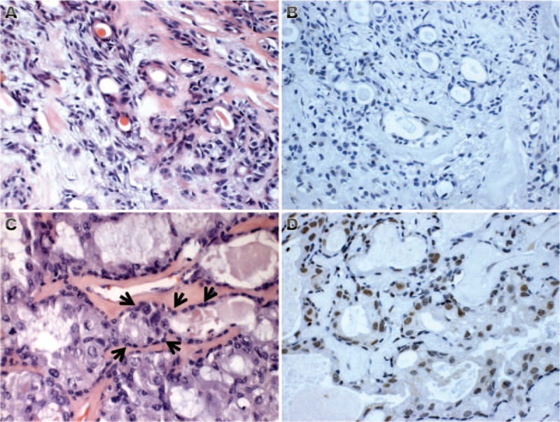
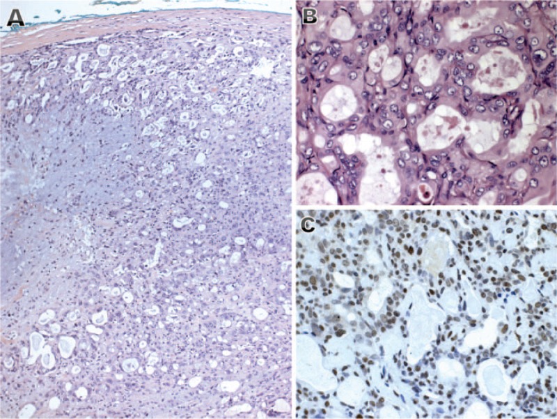
In the present study, we evaluated and described the sensitivity of the stem cell marker B cell-specific moloney murine leukemia virus integration site 1 (Bmi-1) in identifying early lesions of carcinoma ex pleomorphic adenoma (CXPA). While invasive CXPAs are tumors with a prominent and easily recognizable malignant component, the identification of early carcinomatous changes in PA remains a diagnostic challenge due to the lack of objective morphological criteria. The immunohistochemical expression of Bmi-1 was assessed in both adenomatous and carcinomatous components of 9 CXPA cases at an early phase of histological progression (6 intracapsular and 3 minimally invasive) grouped according to the cellular differentiation as luminal (7 cases) or myoepithelial (2 cases). A selective nuclear expression of Bmi-1 was found exclusively in the malignant component of 8 cases (6 luminal type and 2 myoepithelial type), including intraductal carcinoma areas, except for 1 case in which scarce cells of the remnant PA were positive. Thus, Bmi-1 is expressed from the earliest morphologically detectable stages of PA malignant transformation. When faced with atypical features in PA, evaluation of Bmi-1 expression can provide more objective criteria for identification and diagnosis of early lesions of CXPA. This is applied to carcinomas with luminal or myoepithelial differentiation.
Conflict of interest statement
The authors have no conflicts of interest to declare.
Figures

Similar articles
-
Carcinoma ex pleomorphic adenoma (CXPA): immunoprofile of the cells involved in carcinomatous progression.Histopathology. 2005 Jun;46(6):635-41. doi: 10.1111/j.1365-2559.2005.02157.x. Histopathology. 2005. PMID: 15910594
-
Loss of expression of Plag1 in malignant transformation from pleomorphic adenoma to carcinoma ex pleomorphic adenoma.Hum Pathol. 2016 Nov;57:152-159. doi: 10.1016/j.humpath.2016.07.011. Epub 2016 Jul 26. Hum Pathol. 2016. PMID: 27473265
-
Biomarker analysis in carcinoma ex pleomorphic adenoma at an early phase of carcinomatous transformation.Int J Surg Pathol. 2005 Oct;13(4):337-42. doi: 10.1177/106689690501300405. Int J Surg Pathol. 2005. PMID: 16273189
-
Low Grade Carcinoma Ex-Pleomorphic Adenoma: Diagnosis and Diagnostic Challenges Caused by Fine Needle Aspiration: Report of Three Cases and Review of Literature.Head Neck Pathol. 2018 Mar;12(1):82-88. doi: 10.1007/s12105-017-0829-7. Epub 2017 Jun 6. Head Neck Pathol. 2018. PMID: 28589437 Free PMC article. Review.
-
Heterogeneity and versatility of the extracellular matrix during the transition from pleomorphic adenoma to carcinoma ex pleomorphic adenoma: cumulative findings from basic research and new insights.Front Oral Health. 2023 Apr 17;4:942604. doi: 10.3389/froh.2023.942604. eCollection 2023. Front Oral Health. 2023. PMID: 37138857 Free PMC article. Review.
Cited by
-
Primary pulmonary myoepithelial carcinoma in a young woman: A case report and review of literature.Medicine (Baltimore). 2018 Mar;97(9):e0049. doi: 10.1097/MD.0000000000010049. Medicine (Baltimore). 2018. PMID: 29489660 Free PMC article. Review.
-
Nestin Expression Is Associated with Relapses in Head and Neck Lesions.Diagnostics (Basel). 2021 Mar 24;11(4):583. doi: 10.3390/diagnostics11040583. Diagnostics (Basel). 2021. PMID: 33805026 Free PMC article.
-
BMI-1 Expression Heterogeneity in Endometriosis-Related and Non-Endometriotic Ovarian Carcinoma.Int J Mol Sci. 2021 Jun 4;22(11):6082. doi: 10.3390/ijms22116082. Int J Mol Sci. 2021. PMID: 34199929 Free PMC article.
References
-
- Gnepp DR, Brandwein-Gensler MS, El Naggar A. Barnes L, Eveson J, Reichart P, Sidransky D, et al. Carcinoma ex pleomorphic adenoma. World Health Organization. Classification of Tumours. Pathology and Genetics of Head and Neck Tumours. Lyon: IARC Press; 2005. 242–243.
-
- Ellis GL, Auclair PL. Tumors of the Salivary Glands. Washington, DC: Armed Forces Institute of Pathology, ART Press; 2008. 259–269.
-
- Skálová A, Andrle P, Hostička L, et al. Pleomorphic adenoma of salivary glands: diagnostic pitfalls and mimickers of malignancy. Cesk Patol 2012; 48:179–183. - PubMed
-
- Auclair PL, Ellis GL. Atypical features in salivary gland mixed tumors: their relationship to malignant transformation. Mod Pathol 1996; 9:652–657. - PubMed
-
- Takeda Y. An immunohistochemical study of bizarre neoplastic cells in pleomorphic adenoma: its cytological nature and proliferative activity. Pathol Int 1999; 49:993–999. - PubMed
Publication types
MeSH terms
Substances
LinkOut - more resources
Full Text Sources
Medical
