

Multiple Hemolymphangioma of the Visceral Organs: A Case Report and Review of the Literature
- PMID: 26166115
- PMCID: PMC4504602
- DOI: 10.1097/MD.0000000000001126
Multiple Hemolymphangioma of the Visceral Organs: A Case Report and Review of the Literature
Abstract
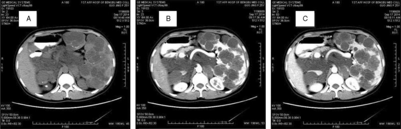
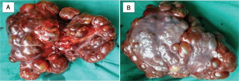
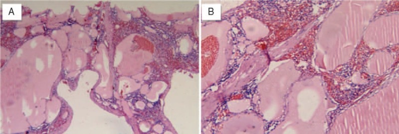
Hemolymphangioma is a rare disease with malformation of both lymphatic and vascular vessels. Few cases of hemolymphangioma occurring in the rectum, small intestine, pancreas, esophagus, and other organs have been reported. Nevertheless, multiple hemolymphangioma of the visceral organs are extremely rare. We report a 25-year-old female with a significantly enlarged spleen full of multiple-rounded lesions. Curiously, the splenic flexure and even retroperitoneum had many lesions. The patient recovered well after splenectomy and the pathologic diagnosis of spleen was hemolymphangioma with abnormal lymphatic and blood vessels with polycystic spaces.Usually, it is hard to cure this disease. We should take much more consideration into the diagnosis, treatment, and even pathogenesis, even though it is a benign lesion.
Conflict of interest statement
The authors have no conflicts of interest to disclose.
Figures
References
-
- Nataf P, Mestiri T, Martin de Lasalle E, et al. Pericardial hemolymphangioma Apropos of a case. Arch Mal Coeur Vaiss 1988; 81:1137–1140. - PubMed
Publication types
MeSH terms
LinkOut - more resources
Full Text Sources
