. 2015 Jul;94(27):e1139.
doi: 10.1097/MD.0000000000001139.

Retrograde Lymphatic Spread of Esophageal Cancer: A Case Report

Affiliations

Retrograde Lymphatic Spread of Esophageal Cancer: A Case Report

Hisashi Oshiro et al. Medicine (Baltimore). 2015 Jul.

Abstract

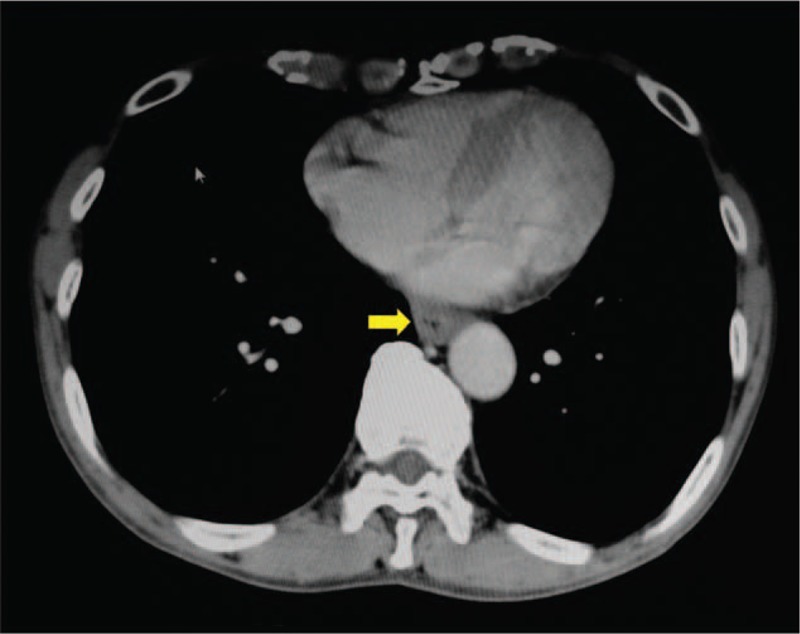
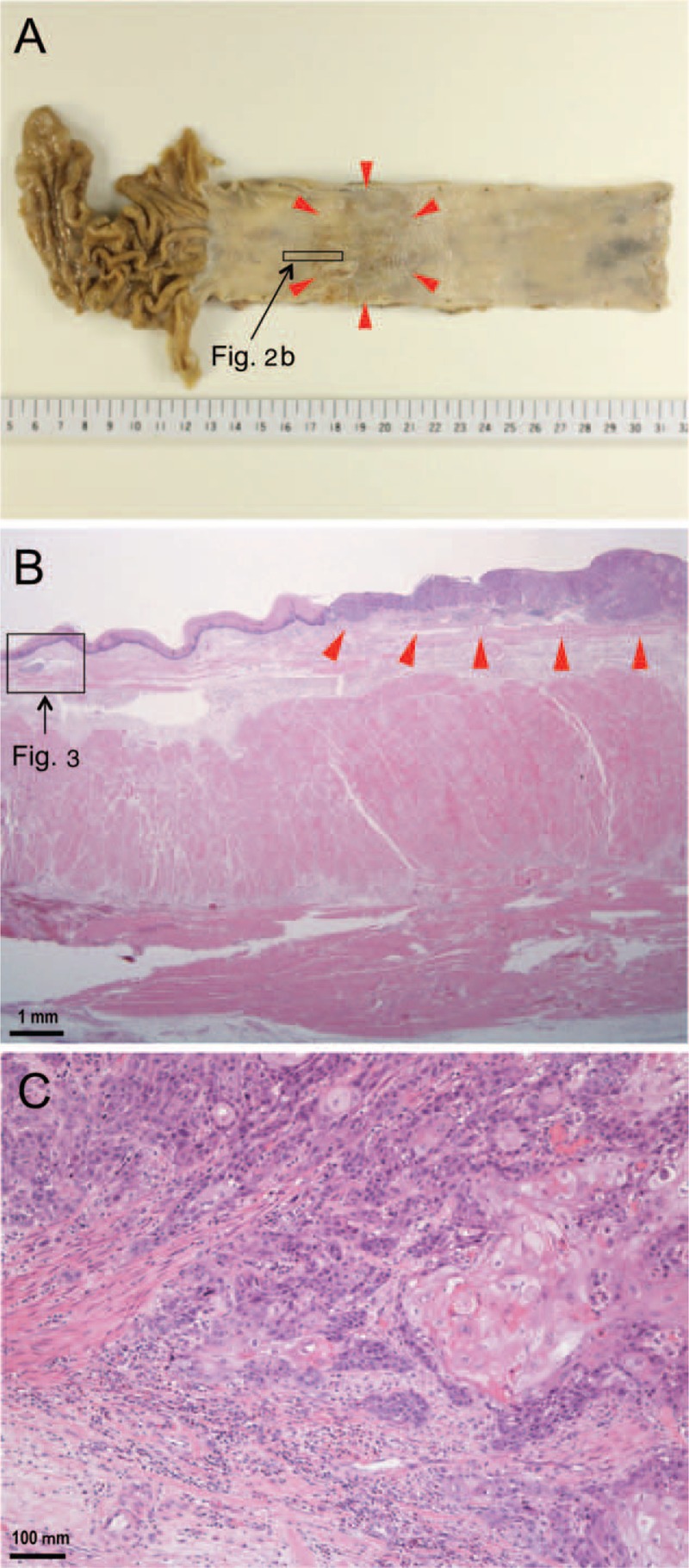
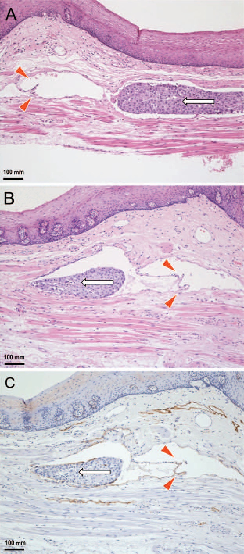
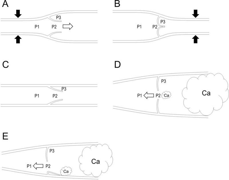
The concept of the retrograde lymphatic spread of cancer cells appears to account for a subset of the essential mechanisms of cancer metastasis in various organs. However, no adequate data currently exist to illustrate the pathology of the retrograde lymphatic metastasis of cancer cells in human bodies. To shed light on this phenomenon, we report a case of a 63-year-old Japanese man who underwent an esophagectomy and lymph node dissection for early-stage esophageal cancer.The patient's clinical information was evaluated by board-certified surgeons and internists. Surgically excised materials were histopathologically evaluated by attending pathologists.Postoperative pathological examination revealed that the patient's tumor was a well-differentiated squamous cell carcinoma with negative surgical margins (T1N0M0, stage I). Apart from the primary lesion, a single lymphatic vessel invasion was found between the lamina propria and lamina muscularis of the esophagus where intralymphatic cancer cells had spread against the direction of backflow prevention valves and skipped beyond these valves without destroying them.The present case demonstrated that the retrograde lymphatic spread of cancer cells can occur in valve-equipped lymphatic vessels. Our study may not only provide a scientific basis for the concept of retrograde lymphatic metastasis but also explain a portion of the complexities associated with the lymphogenous metastasis of esophageal cancer.


Conflict of interest statement

The authors have no conflicts of interest to disclose.

Figures

FIGURE 1
FIGURE 1
A contrast enhanced computed tomographic image of the thoracic organs demonstrates no remarkable changes, except for slight thickening of the wall in the lower thoracic portion of the esophagus (arrow).
FIGURE 2
FIGURE 2
Pathological findings for the examined esophageal cancer. A: A gross image depicts a superficial (0 – Ip + IIc type) single tumor 47 × 38 mm in size located in the middle to the lower thoracic portion of the esophagus (arrowheads). B: A low-power photomicrograph reveals a primary lesion (arrowheads) and an area of retrograde lymphatic invasion distant from the primary lesion (rectangle) (hematoxylin and eosin stain). C: A high-power photomicrograph illustrates the primary lesion and features indicating the invasion of well-differentiated squamous cell carcinoma into the submucosal layer (hematoxylin and eosin stain).
FIGURE 3
FIGURE 3
Histopathological findings regarding the retrograde lymphatic spread of esophageal cancer cells (enlargement of the rectangular area in Figure 2B). A: Distant from the primary lesion, a single lymphatic vessel running along the lamina muscularis has been invaded by cancer cells. Intralymphatic cancer cells (arrow) are spread against the direction of backflow prevention valves (arrowheads). B: A serial tissue section reveals that intralymphatic cancer cells (arrow) skipped beyond the valves (arrowheads) without morphological destruction of the valves (hematoxylin and eosin stain). C: The endothelium is positive for D2-40, demonstrating that the depicted tissue sample is indeed a lymphatic vessel that has been invaded by cancer cells (arrow) (immunohistochemistry).
FIGURE 4
FIGURE 4
Schematic illustrations of lymph flows and fluid pressures at various points in a lymphatic vessel. The labeled pressures are in front of the valve (P1), in the funnel-shaped space between valve faces (P2), and in the pocket (P3). White arrows indicate the flow direction. Black arrows indicate an intrinsic or extrinsic lymphatic pump force. Ca indicates cancer cells. A: If P1 > P2 > P3, the valve is open, and fluid flows in an anterograde direction. B: If P1 = P2 < P3, the valve is closed. C: If P1 = P2 = P3, the stoppage of fluid flow occurs. D: If P1 < P2 = P3, the valve is open, and fluid flows in a retrograde direction. This situation can arise if P3 increases due to the lymphatic invasion of cancer cells and the regurgitation of lymphatic valves occurs due to lymphatic ectasia even if this regurgitation is not continuous. E: If P1 < P2 = P3, the valve is open, and fluid flows in a retrograde direction. This situation can arise if P3 increases due to the lymphatic invasion of cancer cells and the regurgitation of lymphatic valves occurs due to the adhesion of cancer cells to the endothelium even if this regurgitation is not continuous.

References

    1. Akiyama H, Tsurumaru M, Udagawa H, et al. Radical lymph node dissection for cancer of the thoracic esophagus. Ann Surg 1994; 220:364–372. - PMC - PubMed
    1. Ding X, Zhang J, Li B, et al. A meta-analysis of lymph node metastasis rate for patients with thoracic oesophageal cancer and its implication in delineation of clinical target volume for radiation therapy. Br J Radiol 2012; 85:e1110–e1119. - PMC - PubMed
    1. Debois JM. TxNxM1. The Anatomy and Clinics of Metastatic Cancer. New York: Kluwer Academic Publisher; 2002.
    1. Bussani R, De-Giorgio F, Abbate A, et al. Cardiac metastases. J Clin Pathol 2007; 60:27–34. - PMC - PubMed
    1. Leijte JA, van der Ploeg IM, Valdes Olmos RA, et al. Visualization of tumor blockage and rerouting of lymphatic drainage in penile cancer patients by use of SPECT/CT. J Nucl Med 2009; 50:364–367. - PubMed

Publication types