Case Reports
. 2015 Aug 6;97(2):311-8.
doi: 10.1016/j.ajhg.2015.06.003. Epub 2015 Jul 9.

Mutations in KIAA0586 Cause Lethal Ciliopathies Ranging from a Hydrolethalus Phenotype to Short-Rib Polydactyly Syndrome

Affiliations
Case Reports

Mutations in KIAA0586 Cause Lethal Ciliopathies Ranging from a Hydrolethalus Phenotype to Short-Rib Polydactyly Syndrome

Caroline Alby et al. Am J Hum Genet. .

Abstract

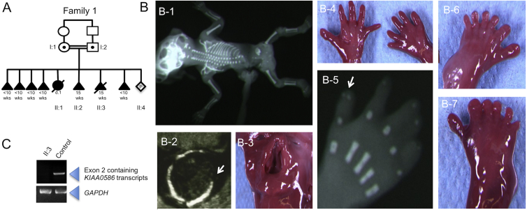
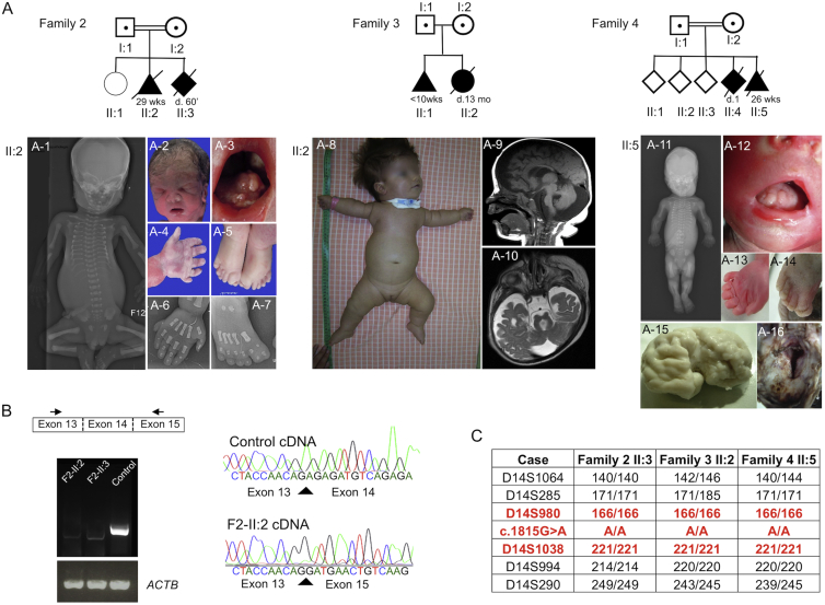
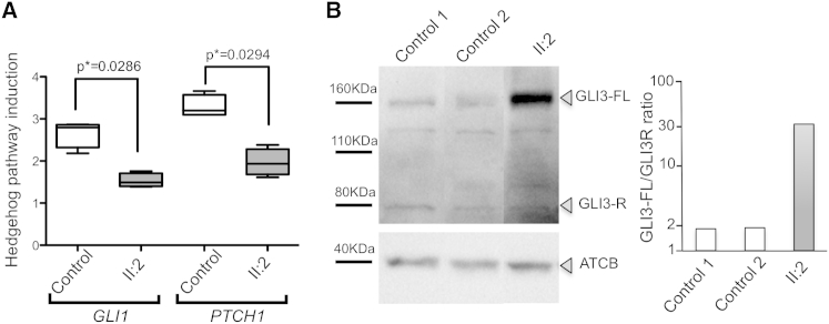
KIAA0586, the human ortholog of chicken TALPID3, is a centrosomal protein that is essential for primary ciliogenesis. Its disruption in animal models causes defects attributed to abnormal hedgehog signaling; these defects include polydactyly and abnormal dorsoventral patterning of the neural tube. Here, we report homozygous mutations of KIAA0586 in four families affected by lethal ciliopathies ranging from a hydrolethalus phenotype to short-rib polydactyly. We show defective ciliogenesis, as well as abnormal response to SHH-signaling activation in cells derived from affected individuals, consistent with a role of KIAA0586 in primary cilia biogenesis. Whereas centriolar maturation seemed unaffected in mutant cells, we observed an abnormal extended pattern of CEP290, a centriolar satellite protein previously associated with ciliopathies. Our data show the crucial role of KIAA0586 in human primary ciliogenesis and subsequent abnormal hedgehog signaling through abnormal GLI3 processing. Our results thus establish that KIAA0586 mutations cause lethal ciliopathies.


Figures

Figure 1
Figure 1
Pedigree and Phenotype of Family 1, Subject II:3 with a KIAA0586 Nonsense Variation (A) Family 1 pedigree. The c.230C>G (p.Ser77) variant segregated with the expected patterns of autosomal-recessive inheritance in all available family members. (B) Phenotype of affected subject II:3. A fetal X-ray shows a frontal view (B-1). Ultrasound imaging shows exencephaly (axial view) with an occipital defect (arrow; B-2). Photographs show cleft lip and palate (B-3), polysyndactyly (B-4) with duplication of the second phalange of thumbs on X-ray of the right hand (arrow; B-5), and preaxial polydactyly of the feet (B-6 and B-7). (C) RT-PCR amplification on mRNA extracted from affected and age-matched-control tissue; amplification shows complete degradation of transcripts containing KIAA0586 exon 2 in mutant tissue. GAPDH was used as cDNA quality and quantity control.
Figure 2
Figure 2
Pedigree, Phenotype, and Haplotype Analyses in Families 2–4 Lead to the Identification of a KIAA0586 Homozygous Splice-Site Mutation with a Founder Effect (A) Pedigrees. In subject II:2 of family 2, an X-ray (frontal view) shows shortening of ribs and micromelia with round metaphysal ends (A-1), and pictures and X-rays show dysmorphism (A-2), lingual hamartomas (A-3), postaxial polydactyly of the hand (A-4 and A-6), and preaxial polysyndactyly of the feet (A-5 and A-7). For subject II:2 in family 3, a picture shows short thorax and micromelia (A-8), and sagittal (A-9) and axial (A-10) views of brain MRI show a micro-brain with large ventricles and large subarachnoid spaces, corpus-callosum and ponto-cerebellar hypoplasia with a large fourth ventricule and cisterna magna, and a molar tooth aspect. In subject II:5 from family 4, an X-ray (frontal view) shows shortening of ribs and micromelia with round metaphysal ends (A-11), and pictures show lingual hamartomas (A-12), postaxial polydactyly of the right hand (A-13), preaxial polydactyly of the right foot (A-14), temporal polymicrogyria (A-15), and an occipital keyhole defect (A-16). (B) The c.1815G>A KIAA0586 variant affects the last base of exon 14 and is responsible for aberrant transcript lacking exon 14, as shown by RT-PCR and sequencing of KIAA0586 mRNA from control and affected subjects (family 2, subjects II:2 and II:3). Total RNAs were extracted from frozen blood with the Nucleospin RNA Blood Kit and on-column DNase digestion (Macherey Nagel). ACTB was used as cDNA quality and quantity control. (C) Haplotype at KIAA0586 of affected subjects from families 2–4.
Figure 3
Figure 3
Analysis of Ciliogenesis in KIAA0586 Mutant Cells Cells from affected individuals were obtained from subject II:2 in family 2. (A and B) In control cells, primary cilia co-stained with acetylated alpha tubulin (ac-α-Tub, clone 6-11B-1, Sigma) and pericentrin (PCNT, Abcam Ab4448) protrude from 60% of cells (n = 500, three independent experiments), whereas only 20% of mutant cells (n = 500) bear a primary cilia. The graph in (B) is a boxplot representing the distribution of the data. The boxes contain 50% of the values and the whiskers indicate the minimum and the maximum of the distribution. The p values were calculated with the Mann-Whitney two-tailed test. Confocal images were taken with a Leica SP5 confocal microscope. (C and D) High-resolution imaging of control and mutant cell centrioles co-stained with ODF2 (centriolar subdistal appendage protein; Abnova, H00004957-M01) and CEP164 (a centriolar distal appendage protein; Sigma, HPA037606) showed no defect in mutant cells, neither on side view (C) nor on top view (D). Super-resolution microscopy was performed with a Leica TCS SP8 STED (Stimulated Emission Depletion). (E) Immunostaining with CEP290 (Novus Biologicals, NB100-86991) and centrin (Clone 20H5, Millipore) antibodies, showing abnormal extended pattern of CEP290.
Figure 4
Figure 4
SHH Signaling in KIAA0586 Mutant Cells (A) KIAA0586 mutant fibroblasts from subject II:2 in family 2 showed an altered response to smoothened agonist (SAG; sc-202814, Santa Cruz; 5 μM for 18 hr), given that they induced less GLI1 and PTCH1 expression than did control cells. Primers used for real-time qRT-PCR are listed in Table S2. Values from five independent experiments were normalized to GAPDH and are presented as relative expression levels. The boxes contain 50% of the values and the whiskers indicate the minimum and the maximum of the distribution. The p values were calculated with the Mann-Whitney two-tailed test. (B) Western blot analysis with a GLI3 antibody (AF3690, R&D Systems) showed that the amount of processing of GLI3-FL into its repressor form, GLI3-R, was much lower in KIAA0586 mutant fibroblasts than in control cells. The samples were not run in contiguous lanes, and in the figure presented here, lanes were spliced together (the entire photograph of the immunoblot is presented in Figure S3). A graphical evaluation of the GLI3-FL/GLI3-R ratio, with actin as a loading control and ImageJ software for densitometry, is presented.

Similar articles

Cited by

References

    1. Huber C., Cormier-Daire V. Ciliary disorder of the skeleton. Am. J. Med. Genet. C. Semin. Med. Genet. 2012;160C:165–174. - PubMed
    1. Putoux A., Thomas S., Coene K.L.M., Davis E.E., Alanay Y., Ogur G., Uz E., Buzas D., Gomes C., Patrier S. KIF7 mutations cause fetal hydrolethalus and acrocallosal syndromes. Nat. Genet. 2011;43:601–606. - PMC - PubMed
    1. Baala L., Romano S., Khaddour R., Saunier S., Smith U.M., Audollent S., Ozilou C., Faivre L., Laurent N., Foliguet B. The Meckel-Gruber syndrome gene, MKS3, is mutated in Joubert syndrome. Am. J. Hum. Genet. 2007;80:186–194. - PMC - PubMed
    1. Delous M., Baala L., Salomon R., Laclef C., Vierkotten J., Tory K., Golzio C., Lacoste T., Besse L., Ozilou C. The ciliary gene RPGRIP1L is mutated in cerebello-oculo-renal syndrome (Joubert syndrome type B) and Meckel syndrome. Nat. Genet. 2007;39:875–881. - PubMed
    1. Valente E.M., Logan C.V., Mougou-Zerelli S., Lee J.H., Silhavy J.L., Brancati F., Iannicelli M., Travaglini L., Romani S., Illi B. Mutations in TMEM216 perturb ciliogenesis and cause Joubert, Meckel and related syndromes. Nat. Genet. 2010;42:619–625. - PMC - PubMed

Publication types

Supplementary concepts

LinkOut - more resources