

OTX2 Transcription Factor Controls Regional Patterning within the Medial Ganglionic Eminence and Regional Identity of the Septum
- PMID: 26166575
- PMCID: PMC4512845
- DOI: 10.1016/j.celrep.2015.06.043
OTX2 Transcription Factor Controls Regional Patterning within the Medial Ganglionic Eminence and Regional Identity of the Septum
Abstract
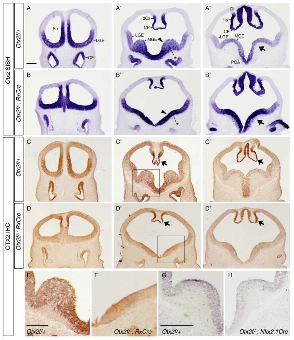
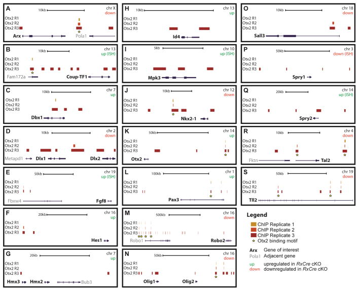
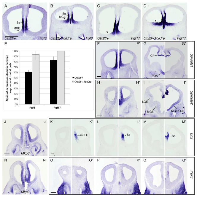
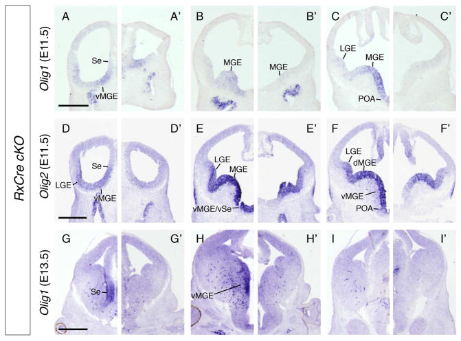
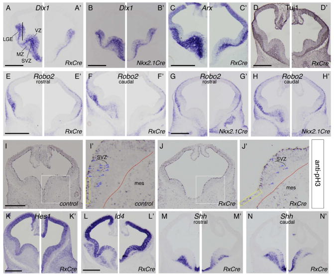
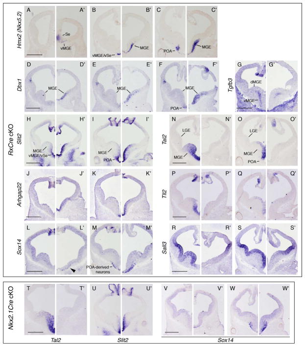
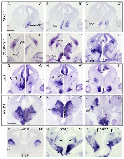
The Otx2 homeodomain transcription factor is essential for gastrulation and early neural development. We generated Otx2 conditional knockout (cKO) mice to investigate its roles in telencephalon development after neurulation (approximately embryonic day 9.0). We conducted transcriptional profiling and in situ hybridization to identify genes de-regulated in Otx2 cKO ventral forebrain. In parallel, we used chromatin immunoprecipitation sequencing to identify enhancer elements, the OTX2 binding motif, and de-regulated genes that are likely direct targets of OTX2 transcriptional regulation. We found that Otx2 was essential in septum specification, regulation of Fgf signaling in the rostral telencephalon, and medial ganglionic eminence (MGE) patterning, neurogenesis, and oligodendrogenesis. Within the MGE, Otx2 was required for ventral, but not dorsal, identity, thus controlling the production of specific MGE derivatives.
Copyright © 2015 The Authors. Published by Elsevier Inc. All rights reserved.
Figures
Similar articles
-
Otx2 and Otx1 protect diencephalon and mesencephalon from caudalization into metencephalon during early brain regionalization.Dev Biol. 2010 Nov 15;347(2):392-403. doi: 10.1016/j.ydbio.2010.08.028. Epub 2010 Sep 16. Dev Biol. 2010. PMID: 20816794
-
Otx2 cell-autonomously determines dorsal mesencephalon versus cerebellum fate independently of isthmic organizing activity.Development. 2014 Jan;141(2):377-88. doi: 10.1242/dev.102954. Epub 2013 Dec 11. Development. 2014. PMID: 24335253
-
Otx2 expression is restricted to dopaminergic neurons of the ventral tegmental area in the adult brain.Int J Dev Biol. 2010;54(5):939-45. doi: 10.1387/ijdb.092974ms. Int J Dev Biol. 2010. PMID: 19924631
-
The homeobox gene Otx2 in development and disease.Exp Eye Res. 2013 Jun;111:9-16. doi: 10.1016/j.exer.2013.03.007. Epub 2013 Mar 21. Exp Eye Res. 2013. PMID: 23523800 Review.
-
The role of Otx2 in organizing the anterior patterning in mouse.Int J Dev Biol. 2001;45(1):337-45. Int J Dev Biol. 2001. PMID: 11291864 Review.
Cited by
-
Transcriptional regulation of MGE progenitor proliferation by PRDM16 controls cortical GABAergic interneuron production.Development. 2020 Nov 16;147(22):dev187526. doi: 10.1242/dev.187526. Development. 2020. PMID: 33060132 Free PMC article.
-
NAT10-mediated ac4C modification promotes ectoderm differentiation of human embryonic stem cells via acetylating NR2F1 mRNA.Cell Prolif. 2024 Apr;57(4):e13577. doi: 10.1111/cpr.13577. Epub 2023 Dec 2. Cell Prolif. 2024. PMID: 38041497 Free PMC article.
-
Tsc1 Loss in VIP-Lineage Cortical Interneurons Results in More VIP+ Interneurons and Enhanced Excitability.Cells. 2023 Dec 26;13(1):52. doi: 10.3390/cells13010052. Cells. 2023. PMID: 38201256 Free PMC article.
-
Neurogenesis From Neural Crest Cells: Molecular Mechanisms in the Formation of Cranial Nerves and Ganglia.Front Cell Dev Biol. 2020 Aug 7;8:635. doi: 10.3389/fcell.2020.00635. eCollection 2020. Front Cell Dev Biol. 2020. PMID: 32850790 Free PMC article. Review.
-
Transcription Factor VAX1 Regulates the Regional Specification of the Subpallium Through Repressing Gsx2.Mol Neurobiol. 2021 Aug;58(8):3729-3744. doi: 10.1007/s12035-021-02378-x. Epub 2021 Apr 5. Mol Neurobiol. 2021. PMID: 33821423
References
-
- Acampora D, Avantaggiato V, Tuorto F, Simeone A. Genetic control of brain morphogenesis through Otx gene dosage requirement. Development. 1997;124(18):3639–50. - PubMed
-
- Acampora D, Mazan S, Lallemand Y, Avantaggiato V, Maury M, Simeone A, Brulet P. Forebrain and midbrain regions are deleted in Otx2−/ − mutants due to a defective anterior neuroectoderm specification during gastrulation. Development. 1995;121(10):3279–90. - PubMed
-
- Acampora D, Simeone A. The TINS Lecture. Understanding the roles of Otx1 and Otx2 in the control of brain morphogenesis. Trends Neurosci. 1999;22(3):116–22. - PubMed
-
- Ang SL, Jin O, Rhinn M, Daigle N, Stevenson L, Rossant J. A targeted mouse Otx2 mutation leads to severe defects in gastrulation and formation of axial mesoderm and to deletion of rostral brain. Development. 1996;122(1):243–52. - PubMed
Publication types
MeSH terms
Substances
Associated data
- Actions
- Actions
- Actions
Grants and funding
- MH049428/MH/NIMH NIH HHS/United States
- MH081880/MH/NIMH NIH HHS/United States
- NS34661/NS/NINDS NIH HHS/United States
- R01 NS034661/NS/NINDS NIH HHS/United States
- F32 MH081431/MH/NIMH NIH HHS/United States
- R01 NS099099/NS/NINDS NIH HHS/United States
- R01 MH081880/MH/NIMH NIH HHS/United States
- T32 HD007470/HD/NICHD NIH HHS/United States
- F32MH081431/MH/NIMH NIH HHS/United States
- R37 MH049428/MH/NIMH NIH HHS/United States
- R01 MH049428/MH/NIMH NIH HHS/United States
- T32 HD 007470/HD/NICHD NIH HHS/United States
LinkOut - more resources
Full Text Sources
Other Literature Sources
Molecular Biology Databases
