. 2015 Jul 13:5:12132.
doi: 10.1038/srep12132.

An anti-inflammatory role for C/EBPδ in human brain pericytes

Affiliations

An anti-inflammatory role for C/EBPδ in human brain pericytes

Justin Rustenhoven et al. Sci Rep. .

Abstract

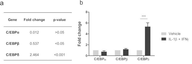
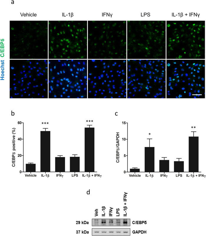
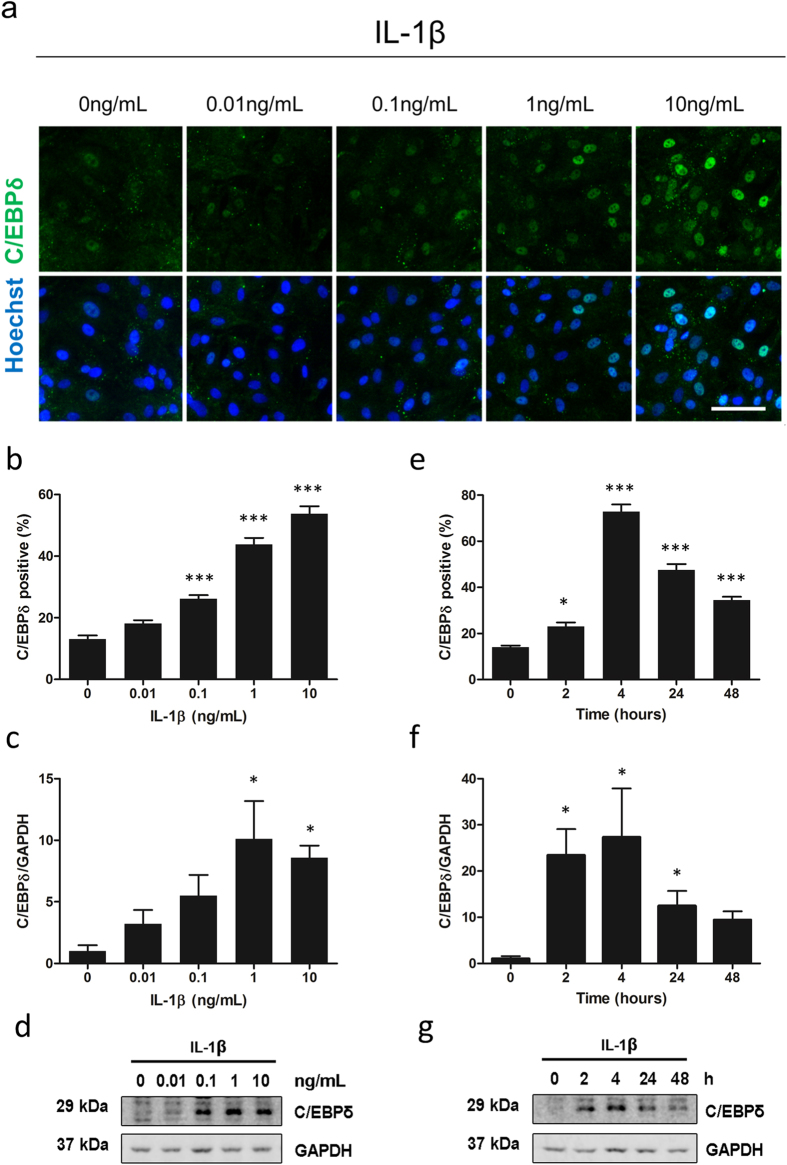
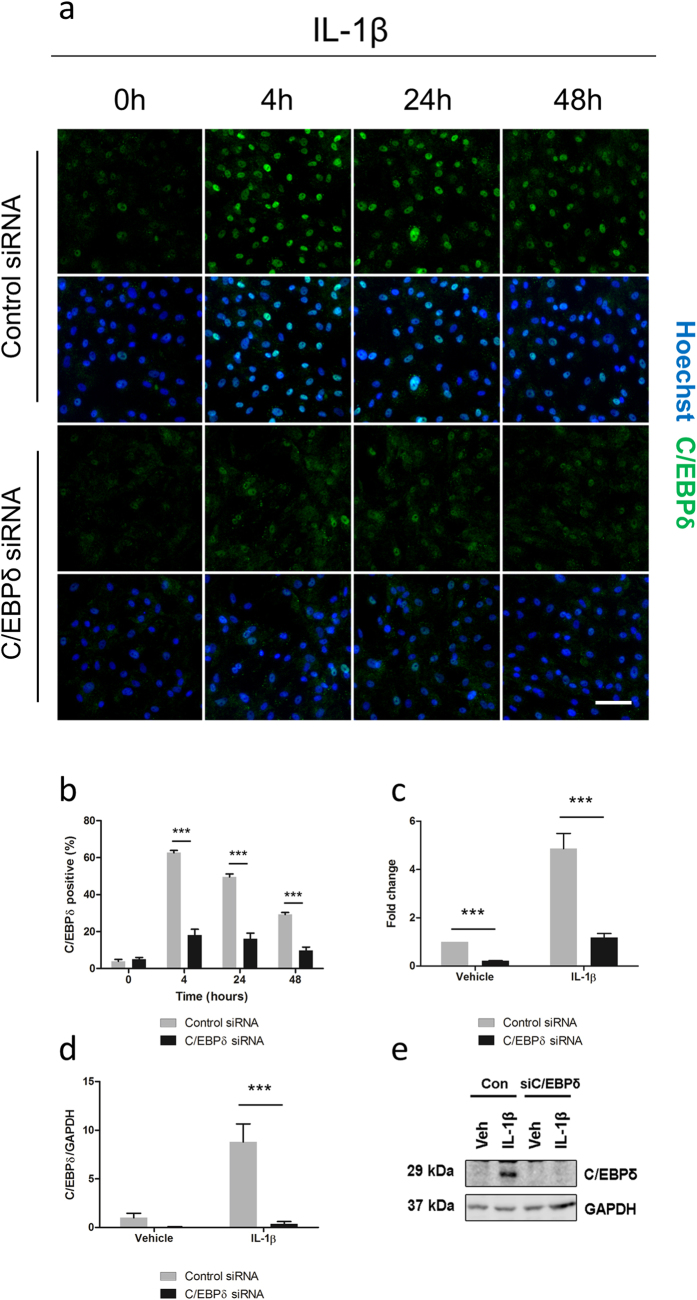
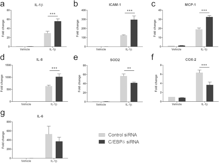
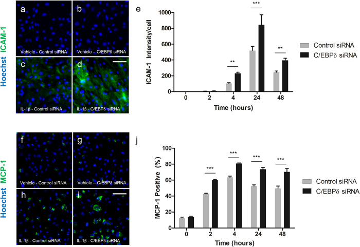
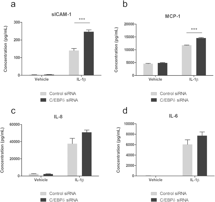
Neuroinflammation contributes to the pathogenesis of several neurological disorders and pericytes are implicated in brain inflammatory processes. Cellular inflammatory responses are orchestrated by transcription factors but information on transcriptional control in pericytes is lacking. Because the transcription factor CCAAT/enhancer binding protein delta (C/EBPδ) is induced in a number of inflammatory brain disorders, we sought to investigate its role in regulating pericyte immune responses. Our results reveal that C/EBPδ is induced in a concentration- and time-dependent fashion in human brain pericytes by interleukin-1β (IL-1β). To investigate the function of the induced C/EBPδ in pericytes we used siRNA to knockdown IL-1β-induced C/EBPδ expression. C/EBPδ knockdown enhanced IL-1β-induced production of intracellular adhesion molecule-1 (ICAM-1), interleukin-8, monocyte chemoattractant protein-1 (MCP-1) and IL-1β, whilst attenuating cyclooxygenase-2 and superoxide dismutase-2 gene expression. Altered ICAM-1 and MCP-1 protein expression were confirmed by cytometric bead array and immunocytochemistry. Our results show that knock-down of C/EBPδ expression in pericytes following immune stimulation increased chemokine and adhesion molecule expression, thus modifying the human brain pericyte inflammatory response. The induction of C/EBPδ following immune stimulation may act to limit infiltration of peripheral immune cells, thereby preventing further inflammatory responses in the brain.


Figures

Figure 1
Figure 1. Characterisation of adult human brain pericyte cultures.
Primary human brain cell cultures at passage five were stained for Hoechst (blue) and cell specific markers αSMA (a), PDGFRβ (b), NG2 (c), P4H (d), Fibronectin (e), CD45 and GFAP (f). Positive controls of astrocytes (GFAP; g) and microglia (CD45; h) at passage two are included. Scale bar = 50 μm.
Figure 2
Figure 2. C/EBPδ is induced in human brain pericytes by IL-1β/IFNγ.
Human brain pericytes were treated with vehicle or 10 ng/mL IL-1β + IFNγ for 24 hours and RNA was extracted. Expression of C/EBPα, C/EBPβ and C/EBPδ was determined by microarray (a) and qRT-PCR (b). Data is displayed as average fold change of five independent cases (a) or mean ± SEM of a separate three independent cases (b). *** = p < 0.001.
Figure 3
Figure 3. C/EBPδ is differentially induced by IFNγ, IL-1β and LPS.
Human brain pericytes were treated with vehicle or 10 ng/mL IL-1β, IFNγ, or LPS for 24 hours. Representative immunocytochemistry images of C/EBPδ with treatments are shown (a). The percentage of cells positive for nuclear C/EBPδ was determined by immunocytochemistry (b) and the intensity of C/EBPδ expression was analysed by western blotting (c,d). Blots are cropped to improve clarity. Full-length blots are presented in Supplementary Figure S2. Data is displayed as mean ± SEM of three independent experiments. * = p < 0.05 compared to vehicle control, ** = p < 0.01 compared to vehicle control *** = p < 0.001 compared to vehicle control. Scale bar = 100 μm.
Figure 4
Figure 4. Time-course and concentration-dependant induction of C/EBPδ expression.
Human brain pericytes were treated with vehicle or 0.01–10 ng/mL IL-1β for 24 hours. Representative immunocytochemistry images of C/EBPδ are shown (a). The percentage of cells positive for nuclear C/EBPδ was determined by immunocytochemistry (b) and C/EBPδ intensity was analysed by western blotting (c,d). Blots are cropped to improve clarity. Full-length blots are presented in Supplementary Figure S2. Human brain pericytes were treated with 10 ng/mL IL-1β for 0–48 hours. The percentage of C/EBPδ positive cells was determined by immunocytochemistry (e) and C/EBPδ intensity was analysed by western blotting (f,g). Data is displayed as mean ± SEM from three independent experiments. * = p < 0.05 compared to vehicle control, ** = p < 0.01 compared to vehicle control, *** = p < 0.001 compared to vehicle control. Scale bar = 100 μm.
Figure 5
Figure 5. Knockdown of C/EBPδ using siRNA.
Human brain pericytes were transfected with 50 nM of control or C/EBPδ siRNA for 48 hours. Following transfection, cells were treated with vehicle or 10 ng/mL IL-1β for 4–48 hours. Representative immunocytochemistry images of C/EBPδ with treatments are shown (a) and the percentage of cells positive for nuclear C/EBPδ was quantified (b). RNA was extracted following a six hour treatment with IL-1β and C/EBPδ transcript expression was determined via RT-qPCR (c). Protein was extracted following a 24 hour treatment with IL-1β and C/EBPδ expression determined by western blotting (d,e). Blots are cropped to improve clarity. Full-length blots are presented in Supplementary Figure S2. *** = p < 0.001. Scale bar = 100 μm.
Figure 6
Figure 6. C/EBPδ knockdown modifies IL-1β induced inflammatory gene expression.
Human brain pericytes were transfected with 50 nM of control or C/EBPδ siRNA for 48 hours. Following transfection, cells were treated with vehicle or 10 ng/mL IL-1β for six hours and RNA was extracted. The effect of C/EBPδ knockdown on IL-1β (a), ICAM-1 (b), MCP-1 (c), IL-8(d), SOD2 (e), COX-2 (f) and IL-6 (g) was assayed by qRT-PCR. Data is displayed as mean ± SEM from four independent cases ** = p < 0.01, *** = p < 0.001.
Figure 7
Figure 7. C/EBPδ knockdown enhances IL-1β induced MCP-1 and ICAM-1 protein expression.
Human brain pericytes were transfected with 50 nM of control or C/EBPδ siRNA for 48 hours. Following transfection, cells were treated with vehicle or 10 ng/mL IL-1β for 2–24 hours and cells fixed and immunostained for ICAM-1 and MCP-1. Representative images of ICAM-1 and MCP-1 immunostaining respectively with vehicle and control siRNA (a,f), vehicle with C/EBPδ siRNA (b,g), 24 hours IL-1β with control siRNA (c,h) and 24 hours IL-1β with C/EBPδ siRNA are shown (d,i). Intensity of ICAM-1 staining (e) and the percentage of MCP-1 positive cells (j) were determined. Data is displayed as mean ± SEM from three independent experiments. ** = p < 0.01, *** = p < 0.001. Scale bar = 100 μm.
Figure 8
Figure 8. C/EBPδ knockdown modifies pericyte secretion of inflammatory mediators.
Human brain pericytes were transfected with 50 nM of control or C/EBPδ siRNA for 48 hours. Following transfection cells were treated with vehicle or 10 ng/mL IL-1β for 24 hours and conditioned media collected. Concentration of sICAM-1 (a), MCP-1 (b), IL-8 (c) and IL-6 (d) in media was determined using a multiplex cytometric bead array. Data represent mean ± SEM (n = 4). ***= p < 0.001.

References

    1. Kleen J. K. & Holmes G. L. Brain inflammation initiates seizures. Nat Med 14, 1309–1310, 10.1038/nm1208-1309 (2008). - DOI - PubMed
    1. Morganti-Kossmann M. C., Rancan M., Stahel P. F. & Kossmann T. Inflammatory response in acute traumatic brain injury: a double-edged sword. Current opinion in critical care 8, 101–105 (2002). - PubMed
    1. del Zoppo G. et al. Inflammation and stroke: putative role for cytokines, adhesion molecules and iNOS in brain response to ischemia. Brain Pathol 10, 95–112 (2000). - PMC - PubMed
    1. Glass C. K., Saijo K., Winner B., Marchetto M. C. & Gage F. H. Mechanisms underlying inflammation in neurodegeneration. Cell 140, 918–934, 10.1016/j.cell.2010.02.016 (2010). - DOI - PMC - PubMed
    1. Streit W. J., Mrak R. E. & Griffin W. S. Microglia and neuroinflammation: a pathological perspective. J Neuroinflammation 1, 14, 10.1186/1742-2094-1-14 (2004). - DOI - PMC - PubMed

Publication types

MeSH terms