. 2015 Jul 16;59(2):298-308.
doi: 10.1016/j.molcel.2015.06.011. Epub 2015 Jul 9.

Glutaminolysis and Transferrin Regulate Ferroptosis

Affiliations

Glutaminolysis and Transferrin Regulate Ferroptosis

Minghui Gao et al. Mol Cell. .

Abstract

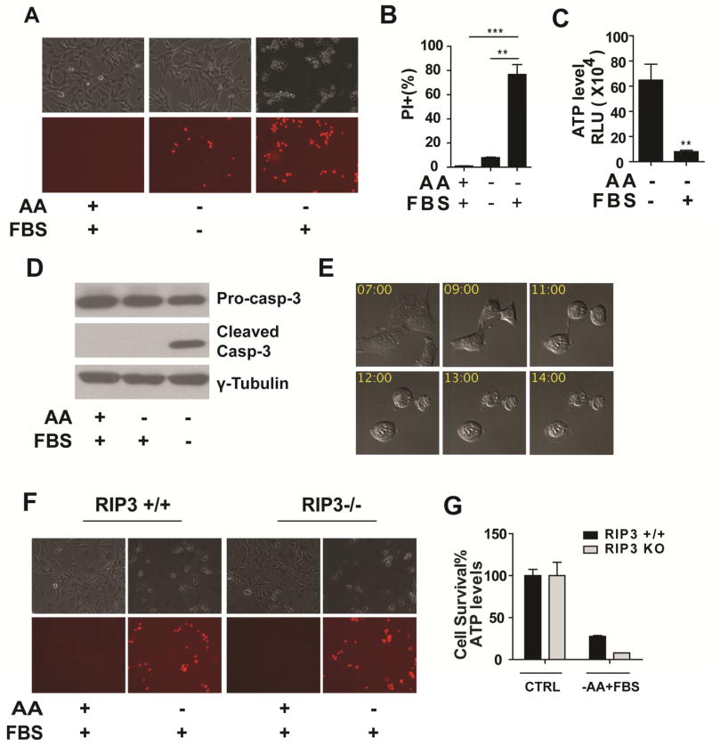
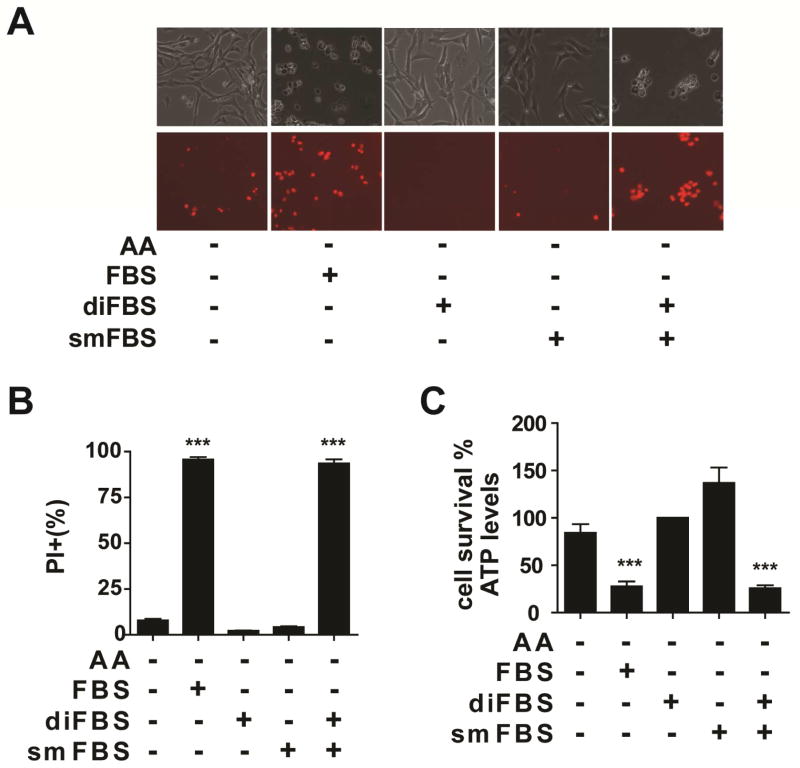
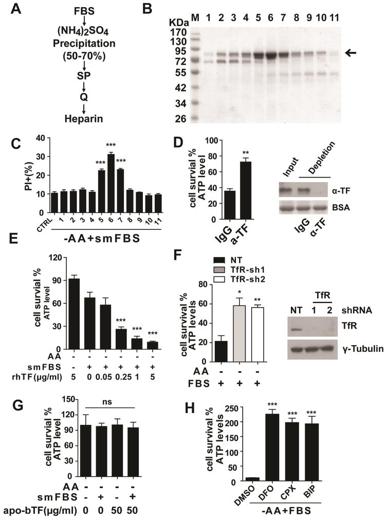
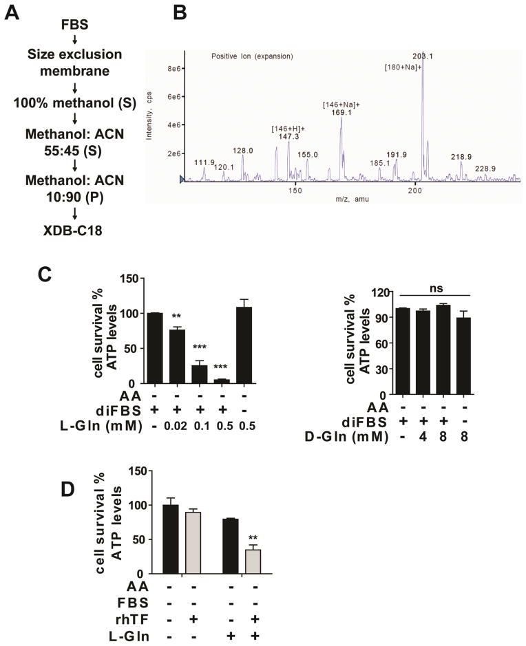
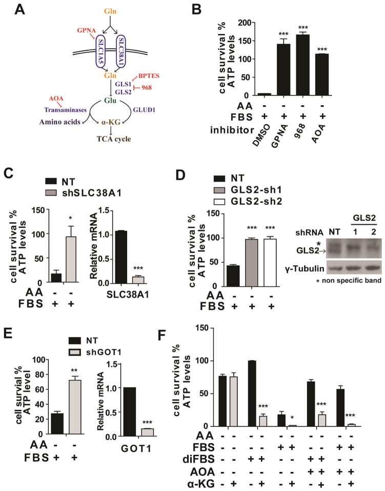
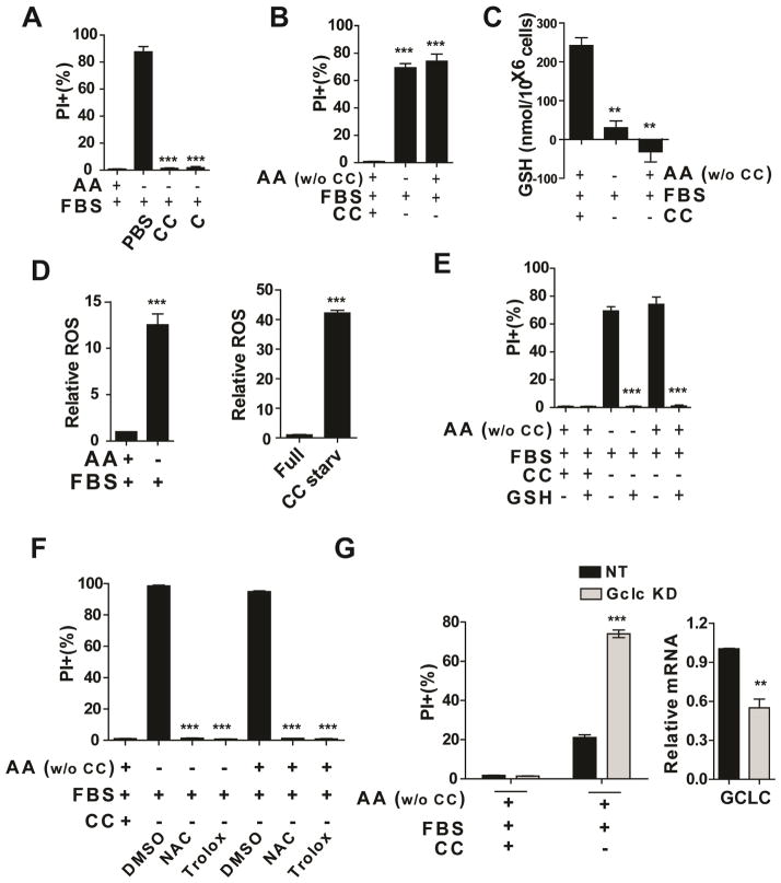
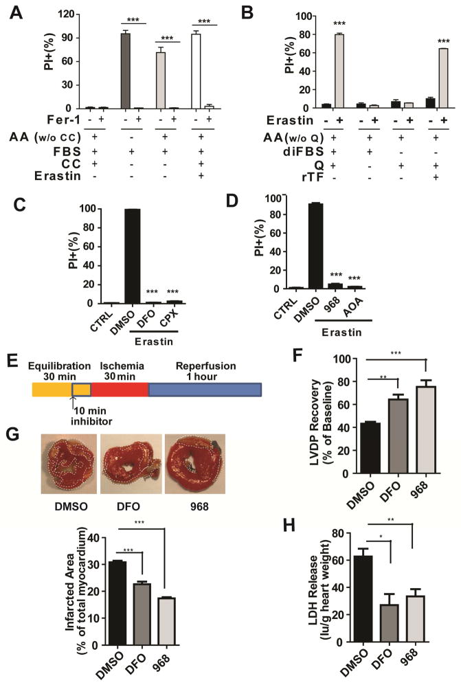
Ferroptosis has emerged as a new form of regulated necrosis that is implicated in various human diseases. However, the mechanisms of ferroptosis are not well defined. This study reports the discovery of multiple molecular components of ferroptosis and its intimate interplay with cellular metabolism and redox machinery. Nutrient starvation often leads to sporadic apoptosis. Strikingly, we found that upon deprivation of amino acids, a more rapid and potent necrosis process can be induced in a serum-dependent manner, which was subsequently determined to be ferroptosis. Two serum factors, the iron-carrier protein transferrin and amino acid glutamine, were identified as the inducers of ferroptosis. We further found that the cell surface transferrin receptor and the glutamine-fueled intracellular metabolic pathway, glutaminolysis, played crucial roles in the death process. Inhibition of glutaminolysis, the essential component of ferroptosis, can reduce heart injury triggered by ischemia/reperfusion, suggesting a potential therapeutic approach for treating related diseases.


Figures

Figure 1
Figure 1. Serum induces potent non-apoptotic, RIP3-indepednent cell death upon amino acid starvation
(A) Microscopy showing cell death induced by serum upon amino acid starvation. MEFs were treated as indicated for 12 hrs. Upper panel: phase-contrast, lower panel: propidium iodide (PI) staining. AA: amino acids, FBS: 10% (v/v) Fetal Bovine Serum. (B) Quantitation of cell death by PI-staining coupled with flow cytometry. Cells were subjected to the same treatment as in panel A. Quantitative data here and thereafter are presented as mean ± SEM from 3 independent experiments (*P < 0.05, **P < 0.01, ***P < 0.001 by unpaired Student t-test). (C) Determination of cell viability by measuring cellular ATP levels. Cells were subjected to the same treatment as in panel A. (D) Serum-induced cell death is independent of caspase activation. MEFs were treated as indicated for 12 hrs and caspase-3 activation was assessed by immunoblotting. (E) Serum-induced cell death shows necrotic morphology. Representative still images from confocal time-lapse imaging of MEFs treated with amino acid starvation in the presence of FBS. Time (hr) after treatment is indicated. (F–G) Serum-induced cell death is independent of RIP3. RIP3+/+ MEFs and RIP3−/− MEFs were treated as indicated. Cell death was monitored by phase-contract microscopy and PI-staining (F), and cell viability was determined by measuring cellular ATP levels (G). CTRL: control. See also Figure S1 and Movies S1, S2 and S3
Figure 2
Figure 2. Multiple serum components are required for serum-induced cell death
(A) MEFs were treated as indicated for 12 hrs. Upper panel: phase-contrast, Lower panel: PI staining. diFBS: 10% (v/v) dialyzed FBS, smFBS: 10% (v/v). (B–C) MEFs were treated as indicated for 12 hrs. Cell death was determined by PI staining coupled with flow Cytometry (B), and cell viability was determined by measuring cellular ATP levels (C). See also Figure S2 and Movie S4
Figure 3
Figure 3. Transferrin and transferrin receptor are required for serum-dependent necrosis
(A) Purification scheme for the death-inducing component in diFBS. See Methods for detailed description. (B) The final heparin column fractions were resolved by SDS-PAGE and stained with Coomassie blue. Arrow indicates the protein band correlating with killing activity. (C) In amino acid-free medium, bax/bak-DKO MEFs were incubated with the heparin fractions in combination with smFBS as indicated, and cell death was determined by PI staining coupled with flow cytometry. (D) Immuno-depletion of transferrin (TF) abrogated the killing activity of serum. Serum was immuno-depleted with control IgG or anti-transferrin antibody (α-TF) as indicated, and subsequently used to induce cell death in Bax/bak-DKO MEFs under amino acid-free conditions for 12 hrs. Western blot showed the efficiency of transferrin depletion from FBS, with BSA as the loading control. (E) Recombinant human holo-transferrin (rhTF) induced cell death in Bax/bak-DKO MEFs under amino acid-free conditions in a smFBS-dependent manner. (F) RNAi of transferrin receptor (TfR) inhibited serum-dependent necrosis. MEFs expressing control shRNA (NT) or two independent shRNAs targeting TfR were treated as indicated for 12 hrs and cell viability was measured. Western blot (lower panel) confirmed knockdown of TfR expression. (G) Iron-free bovine apo-transferrin (apo-bTF) did not have death-inducing activity. (H) Iron chelators blocked serum-dependent necrosis. MEFs were treated with 3 different iron chelators as indicated, and cell viability was determined by measuring cellular ATP levels. DFO (Deferoxamine), 80 μM; CPX (ciclopirox olamine), 10 μM; BIP (2, 2-bipyridyl), 100 μM. See also Figure S3
Figure 4
Figure 4. Glutamine is the death-inducing small molecule component in serum
(A) Purification scheme for the death-inducing small molecule component in FBS. S: supernatant; P: pellet. See Methods for detailed description. (B) LC/MS spectra of the active fraction from the XDB-C18 column. (C) L-Gln (left panel) but not D-Gln (right panel) induces cell death in a diFBS-dependent manner under AA starvation. MEFs were treated as indicated for 12 hrs and cell viability was subsequently measured. (D) L-Gln in combination with transferrin recapitulated the cell death-inducing activity of serum. bax/bak-DKO MEFs were treated as indicated for 12 hrs and cell viability was subsequently measured. L-Gln concentration, 0.1 mM. See also Figure S4
Figure 5
Figure 5. Glutaminolysis mediates serum-dependent necrosis
(A) Schematic overview of the glutaminolysis pathway. (B) Pharmacological inhibition of multiple components in the glutaminolysis pathway abrogated serum-dependent necrosis. The following inhibitors were used as indicated: L-Gln transporter inhibitor GPNA (5 mM); GLS inhibitor compound 968 (968, 20 μM); Pan-transaminases inhibitor AOA (0.5 mM). (C) RNAi knockdown of SLC38A1 inhibited serum-dependent necrosis. Left: MEFs expressing non-targeting (NT) shRNA or shRNA targeting SLC38A1 were treated as indicated for 12 hrs and cell viability was subsequently measured. Right: qPCR measurement of SLC38A1 mRNA levels in MEFs infected with NT shRNA or shRNA targeting SLC38A1. (D) Knockdown of GLS2 blocked serum-dependent necrosis. MEFs expressing non-targeting shRNA (NT) or two independent shRNAs targeting GLS2 were treated as indicated for 12 hrs and cell viability was subsequently measured. FBS: 5% (v/v). Western blotting (lower panel) confirmed the knockdown of GLS2 expression. (E) GOT1 RNAi reduced serum-dependent necrosis. Left: MEFs expressing non-targeting (NT) shRNA or shRNA targeting GOT1 were treated as indicated for 12 hrs and cell viability was subsequently measured. Right: qPCR measurement of GOT1 mRNA levels in MEFs infected with NT shRNA or shRNA targeting GOT1. (F) α-ketoglutarate can mimic the death-inducing activity of L-Gln but in a manner insensitive to the transaminase inhibitor AOA. MEFs were treated as indicated for 12 hrs, and cell viability was subsequently measured. α-KG: (Dimethyl-α-Ketoglutarate), 4 mM; AOA, 0.5 mM. See also Figure S5
Figure 6
Figure 6. Cystine starvation and subsequent cellular redox homeostasis unbalance trigger serum-dependent necrosis
(A) Addition of cysteine (C, 0.2 mM) or cystine (CC, 0.2 mM) inhibited serum-dependent necrosis. MEFs were treated as indicated for 12 hrs. Cell death was subsequently measured by PI staining followed by flow cytometry. (B) Cystine starvation alone is sufficient to induce cell death. MEFs were treated as indicated for 12 hrs and cell death was determined by PI staining coupled with flow cytometry. (C) Glutathione (GSH) was depleted in the condition of serum-induced necrosis or cystine starvation. MEFs were treated as indicated for 4 hrs and harvested for total glutathione measurement. (D) Accumulation of ROS in MEFs induced by serum under AA starvation condition (left) or cystine starvation (right). ROS levels in MEFs were determined by H2DCFDA staining. H2DCFDA assay was performed 8 hrs (Left) or 6 hours (right). (E) Supplement of GSH blocked cell death induced by serum upon total AA starvation or cystine starvation. MEFs were treated as indicated for 12 hrs, and cell death was subsequently measured. (F) Cell death induced by serum upon AA starvation or cystine starvation can be prevented by antioxidant reagents NAC (0.2 mM) and Trolox (0.2 mM). MEFs were treated as indicated for 12 hrs, and cell death was subsequently measured. (G) GCLC RNAi sensitized serum-dependent cell death upon cystine starvation. Left: qPCR measurement of GCLC mRNA levels in MEFs infected with NT shRNA or shRNA targeting GCLC. Right: MEFs expressing non-targeting (NT) shRNA or shRNA targeting GCLC were treated as indicated for 10 hrs and cell death was subsequently measured. See also Figure S6
Figure 7
Figure 7. Serum-dependent necrosis is ferroposis and is involved in ischemia-reperfusion heart injury
(A) Ferroptosis inhibitor Ferrastatin-1 (Fer-1) can inhibit serum-induced necrosis upon total AA starvation or cystine (CC) starvation. MEFs were treated as indicated for 12 hrs, and cell viability was subsequently measured by PI staining followed by flow cytometry. Erastin: 1 μM. (B) Erastin-induced ferroptosis required both transferrin and glutamine. bax/bak DKO MEFs were treated as indicated for 12 hrs, and cell death was subsequently measured by PI staining followed by flow cytometry. Erastin: 1 μM. (C) Iron chelators inhibited erastin-induced ferroptosis. MEFs were treated as indicated for 12 hrs, and cell death was subsequently measured. DFO, 80 μM; CPX, 10 μM; Erastin: 1 μM. (D) Inhibition of GLS by compound 968 (20 μM) or transaminases by pan-transaminases inhibitor AOA (0.5 mM) blocked erastin-induced ferroptosis. MEFs were treated as indicated for 12 hrs, and cell death was subsequently measured. Erastin, 1 μM. (E) Schematic protocol for the ischemia-reperfusion study involving 30-min of global ischemia followed by a 60-min reperfusion period. (F) Determination of myocardial ischemic injury and function by left ventricular developed pressure (LVDP) recovery, showing improved functional recovery upon treatment with DFO or compound-968 (968). DFO: 80 μM, 968: 25 μM. (G) Representative cross-sections of hearts stained with TTC, demonstrating reduced infarct injury (pale region) upon DFO or 968 treatment. The plot (lower panel) shows quantification of infarct size of each group. (H) Determination of myocardial ischemic injury by lactate dehydrogenase (LDH) release, demonstrating reduced infarct injury in the hearts treated with DFO or 968. See also Figure S7

References

    1. Ananthakrishnan R, Kaneko M, Hwang YC, Quadri N, Gomez T, Li Q, Caspersen C, Ramasamy R. Aldose reductase mediates myocardial ischemia-reperfusion injury in part by opening mitochondrial permeability transition pore. American journal of physiology Heart and circulatory physiology. 2009;296:H333–341. - PMC - PubMed
    1. Andrews NC, Schmidt PJ. Iron homeostasis. Annual review of physiology. 2007;69:69–85. - PubMed
    1. Bergsbaken T, Fink SL, Cookson BT. Pyroptosis: host cell death and inflammation. Nat Rev Microbiol. 2009;7:99–109. - PMC - PubMed
    1. Beutler E, Gelbart T, Kondo T, Matsunaga AT. The molecular basis of a case of gammaglutamylcysteine synthetase deficiency. Blood. 1999;94:2890–2894. - PubMed
    1. Blois MS. Antioxidant Determinations by the Use of a Stable Free Radical. Nature. 1958;181:1199–1200.

Publication types

MeSH terms