. 2009 Jun;34(6):441-448.
doi: 10.1557/mrs2009.120.

Magnetic Nanoparticles for Early Detection of Cancer by Magnetic Resonance Imaging

Magnetic Nanoparticles for Early Detection of Cancer by Magnetic Resonance Imaging

Wenbin Lin et al. MRS Bull. 2009 Jun.

Abstract

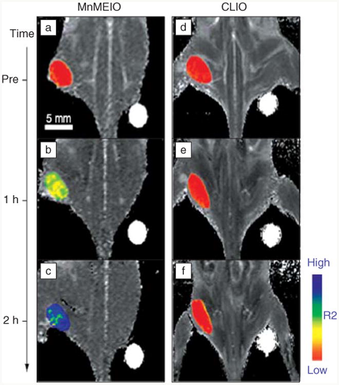
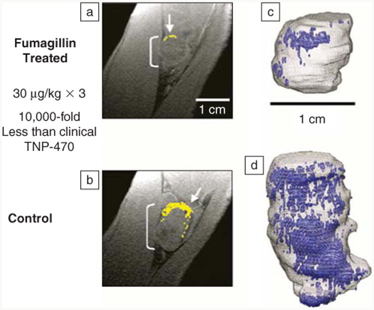
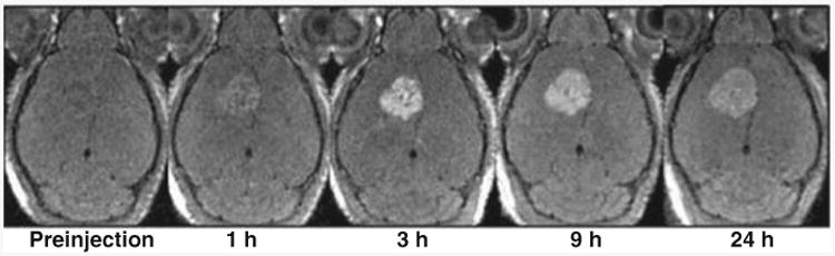
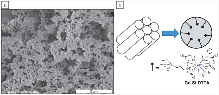
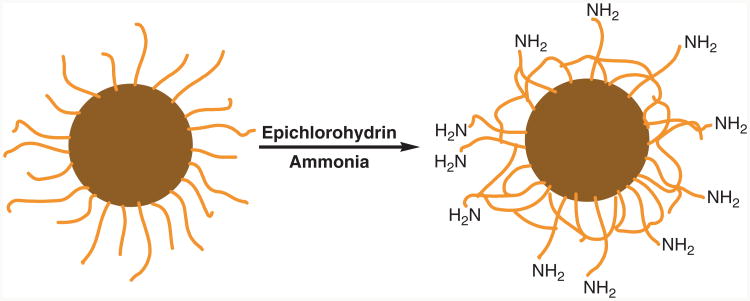
This article provides a brief overview of recent progress in the synthesis and functionalization of magnetic nanoparticles and their applications in the early detection of malignant tumors by magnetic resonance imaging (MRI). The intrinsic low sensitivity of MRI necessitates the use of large quantities of exogenous contrast agents in many imaging studies. Magnetic nanoparticles have recently emerged as highly efficient MRI contrast agents because these nanometer-scale materials can carry high payloads while maintaining the ability to move through physiological systems. Superparamagnetic ferrite nanoparticles (such as iron oxide) provide excellent negative contrast enhancement. Recent refinement of synthetic methodologies has led to ferrite nanoparticles with narrow size distributions and high crystallinity. Target-specific tumor imaging becomes possible through functionalization of ferrite nanoparticles with targeting agents to allow for site-specific accumulation. Nanoparticulate contrast agents capable of positive contrast enhancement have recently been developed in order to overcome the drawbacks of negative contrast enhancement afforded by ferrite nanoparticles. These newly developed magnetic nanoparticles have the potential to enable physicians to diagnose cancer at the earliest stage possible and thus can have an enormous impact on more effective cancer treatment.


Figures

Figure 1
Figure 1
Cross-linked iron oxide (CLIO) nanoparticles for T2-weighted images of rodent pancreatic cancer: (a) preinjection of CLIO, (b) postinjection of CLIO, and (c) higher magnification of postinjection image with the arrow indicating tumor. L, liver; P, pancreas; K, kidney; B, bowel.
Figure 2
Figure 2
In vivo magnetic resonance detection of cancer after administration of magnetic nanoparticles Herceptin conjugates. MnFe2O4 nanoparticles (MnMEIO) (a–c) show higher signal enhancement than cross-linked iron oxide (CLIO) (d–f). R2, inverse of transverse relaxation time.
Figure 3
Figure 3
The synthesis of magnetite nanoparticle/mesoporous silica core (Fe3O4@mSiO2)-shell nanostructures and its in vivo dual modal imaging (magnetic resonance imaging [MRI] and optical imaging). PEG, poly(ethylene glycol); CTAB, cetyl trimethyl ammonium bromide.
Figure 4
Figure 4
Scanning electron microscope image of porcine aortic smooth muscle cells known to overexpress cell surface tissue factor (TF) in culture. (a) Cells exposed to TF-targeted microemulsion nanoparticles. (b) Cells pretreated with anti-tissue factor antibody and subsequently exposed to TF-targeted microemulsion nanoparticles.
Figure 5
Figure 5
(a) Diminished αVβ3-integrin contrast enhancement in a T1-weighted 3D gradient echo magnetic resonance (1.5 T) single slice image of a VX2 rabbit tumor following αVβ3-targeted fumagillin nanoparticles versus (b) those given αVβ3-targeted nanoparticles without drug. (Enhancing pixels are color coded in yellow.) (c) 3D angiogenesis maps in VX2 tumors following αVβ3-targeted fumagillin nanoparticles versus (d) αVβ3-nanoparticles without drug.
Figure 6
Figure 6
Breast cancer cells were selectively enhanced in T1-weighted magnetic resonance imaging by Herceptin-functionalized MnO nanoparticles.
Figure 7
Figure 7
(a) Transmission electron microscopy image and (b) schematic representation of layer-by-layer self-assembled nanoparticles decorated with arginine-glycine-aspartate (RGD) peptides for integrin targeting. (c) T1-weighted magnetic resonance images of HT-29 (human colon cancer) cells that have been incubated with various nanoparticles. From left to right, control cells without any nanoparticle, cells with layer-by-layer nanoparticles, cells with layer-by-layer nanoparticles that were functionalized with RGD peptide, and cells with layer-by-layer nanoparticles that were functionalized with RGD peptide. (d) Phase contrast optical and (e) confocal fluorescence images of HT-29 cells incubated with layer-by-layer nanoparticles that were functionalized with RGD peptide.
Figure 8
Figure 8
(a) Scanning electron microscope image of mesoporous silica nanospheres showing the formation of monodisperse, water-dispersable nanoparticles. (b) Schematic showing the Gd-(trimethoxysilylpropyl)diethylenetriamine tetraacetate (Gd-Si-DTTA) complexes residing in hexagonally ordered nanochannels of ∼2.4 nm in diameter.
Figure 9
Figure 9
Scanning electron microscope images of Gd(BDC)1.5(H2O)2 nanorods synthesized with w = 5 at (a) lower magnficiation and (b) higher magnification; and w = 10 at (c) lower magnification and (d) higher magnification. BDC is 1,4-benzenedicarboxylate. w, water to surfactant molar ratio.
Scheme 1
Scheme 1
Preparation of iron oxide nanoparticles with cross-linked dextran surface coating (light brown strands) and amino functional groups for bioconjugation.
Scheme 2
Scheme 2
Layer-by-layer self-assembly of multifunctional hybrid nanoparticles for an increased Gd payload. PSS, polystyrene sulfonate; DTTA, diethylenetriamine tetraacetate.

References

    1. Stark DD, Bradley WG., Jr . Magnetic Resonance Imaging. Mosby; St. Louis: 1999.
    1. Aime S, Botta M, Fasano M, Terreno E. Chem Soc Rev. 1998;27:19.
    1. Caravan P, Ellison JJ, McMurry TJ, Lauffer RB. Chem Rev. 1999;99:2293. - PubMed
    1. Larson SM. Cancer. 1991;67(Suppl. 4):1253. - PubMed
    1. Yang DJ, Kim EE, Inoue T. Ann Nucl Med. 2006;20:1. - PubMed

LinkOut - more resources