Review
. 2015 Jul-Sep;31(3):185-93.
doi: 10.4103/0970-1591.156924.

Advances in CT imaging for urolithiasis

Affiliations
Review

Advances in CT imaging for urolithiasis

Yasir Andrabi et al. Indian J Urol. 2015 Jul-Sep.

Abstract

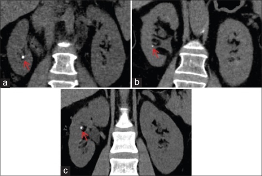
Urolithiasis is a common disease with increasing prevalence worldwide and a lifetime-estimated recurrence risk of over 50%. Imaging plays a critical role in the initial diagnosis, follow-up and urological management of urinary tract stone disease. Unenhanced helical computed tomography (CT) is highly sensitive (>95%) and specific (>96%) in the diagnosis of urolithiasis and is the imaging investigation of choice for the initial assessment of patients with suspected urolithiasis. The emergence of multi-detector CT (MDCT) and technological innovations in CT such as dual-energy CT (DECT) has widened the scope of MDCT in the stone disease management from initial diagnosis to encompass treatment planning and monitoring of treatment success. DECT has been shown to enhance pre-treatment characterization of stone composition in comparison with conventional MDCT and is being increasingly used. Although CT-related radiation dose exposure remains a valid concern, the use of low-dose MDCT protocols and integration of newer iterative reconstruction algorithms into routine CT practice has resulted in a substantial decrease in ionizing radiation exposure. In this review article, our intent is to discuss the role of MDCT in the diagnosis and post-treatment evaluation of urolithiasis and review the impact of emerging CT technologies such as dual energy in clinical practice.

Keywords: Advances; computed tomography; urolithiasis.


Conflict of interest statement

Conflict of Interest: None declared.

Figures

Figure 1
Figure 1
Volumetric assessment of renal stone burden. Axial non-contrast computed tomography image obtained prior to percutaneous nephrolithotomy demonstrates region of interest-based segmentation method for volume estimation
Figure 2
Figure 2
Characterization of kidney stones using dual-energy computed tomography (DECT). DECT helps to distinguish between uric acid and non-uric acid renal stones. a and c are axial images acquired from different patients who presented with flank pain. (a) Axial image showing a non-uric acid renal stone in the right kidney (arrowhead) colored in blue. (b) Graph showing the composition of this stone (blue arrowhead). (c) Axial image showing a uric acid renal calculus in the left kidney (arrow) colored in red. (d) Graph confirms the composition of the stone (red arrowhead)
Figure 3
Figure 3
Measurement of stone-to-skin distance (SSD) in a 54-year-old man. On an axial non-contrast computed tomography scan, the distance from the center of the stone to the surface of the skin at 0°, 45° and 90° is 8.0, 8.7 and 9.4 cm, respectively. The mean of these three measurements is used to represent the average SSD, which is 8.7 cm
Figure 4
Figure 4
Staghorn calculus in a 47-year-old man with treated lithotripsy. (a) Axial non-contrast computed tomography (CT) obtained prior to lithotripsy shows large right staghorn calculus with hydronephrosis of the upper pole renal calyces. (b) Post-treatment CT scan in soft tissue window settings (400/30) shows reduction in stone burden with residual stone fragments in the renal pelvis. (c) CT image in the bone window settings (1100/300) allows improved distinction of the stone from the ureteral stent
Figure 5
Figure 5
Iterative reconstruction techniques in computed tomography (CT) for urolithiasis. Iterative reconstruction (IR) techniques reduce image noise and allow acquisition of CT scans with low radiation dose, preserving diagnostic accuracy. Coronal images from a patient with medullary sponge kidney acquired in different dates. (a) Image reconstructed with Filter back projection (CTDIvol: 11.45 mGy) and (b) image reconstructed with an IR technique (adaptive statistical iterative reconstruction 60%) (CTDIvol: 7.01 mGy). Note the decreased image noise, preserved image quality and visualization of the calculi in the image obtained with the IR technique compared that obtained with Filtered Back Projection (FBP). Also note the radiation dose reduction obtained using the IR technique and different image texture on the images obtained with the IR technique
Figure 6
Figure 6
Iterative reconstruction (IR) levels in computed tomography (CT) scans performed for renal stones. IR techniques decrease image noise, allowing radiation dose reduction, while image quality is preserved or even improved. High levels of IR techniques are desired for CT scans performed for renal stones. (a–c) Coronal images from different patients generated with high levels of different IR techniques, showing renal stones in the right kidney (red arrows). (a) Image reconstructed with adaptive statistical iterative reconstruction 80%; patient's weight 70 kg and CTDIvol: 3.84 mGy. (b) iDose4 L5; patient's weight 71 kg and CTDIvol: 5.66 mGy. (c) Sonogram affirmed iterative reconstruction 4; patient's weight 81 kg and CTDIvol: 8.74 mGy. Note the comparable image quality and contrast preservation among the different images, allowing the diagnosis of urolithiasis

Similar articles

Cited by

References

    1. Neisius A, Preminger GM. Stones in 2012: Epidemiology, prevention and redefining therapeutic standards. Nat Rev Urol. 2013;10:75–7. - PubMed
    1. Bartoletti R, Cai T, Mondaini N, Melone F, Travaglini F, Carini M, et al. Epidemiology and risk factors in urolithiasis. Urol Int. 2007;79(Suppl 1):3–7. - PubMed
    1. Scales CD, Jr, Smith AC, Hanley JM, Saigal CS. Urologic Diseases in America Project. Prevalence of kidney stones in the United States. Eur Urol. 2012;62:160–5. - PMC - PubMed
    1. Seitz C, Fajkovic H. Epidemiological gender-specific aspects in urolithiasis. World J Urol. 2013;31:1087–92. - PubMed
    1. Smith RC, Verga M, McCarthy S, Rosenfield AT. Diagnosis of acute flank pain: Value of unenhanced helical CT. AJR Am J Roentgenol. 1996;166:97–101. - PubMed