. 2015:2015:160454.
doi: 10.1155/2015/160454. Epub 2015 Jun 8.

Advanced Morphological and Functional Magnetic Resonance Techniques in Glaucoma

Affiliations

Advanced Morphological and Functional Magnetic Resonance Techniques in Glaucoma

Rodolfo Mastropasqua et al. Biomed Res Int. 2015.

Abstract

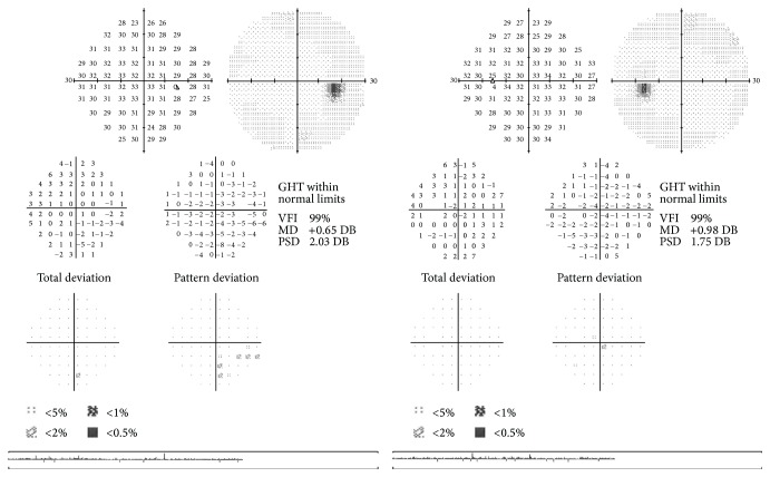
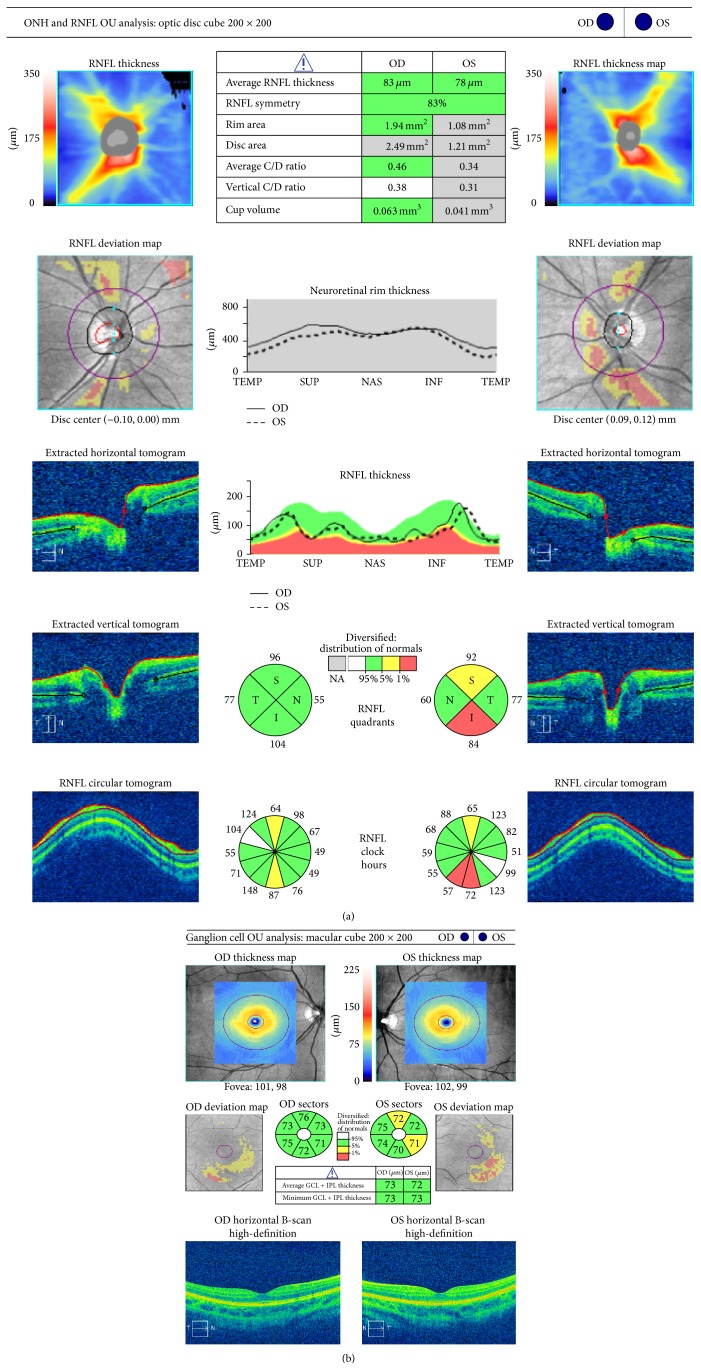
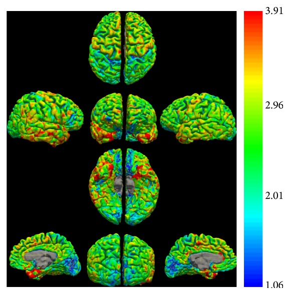
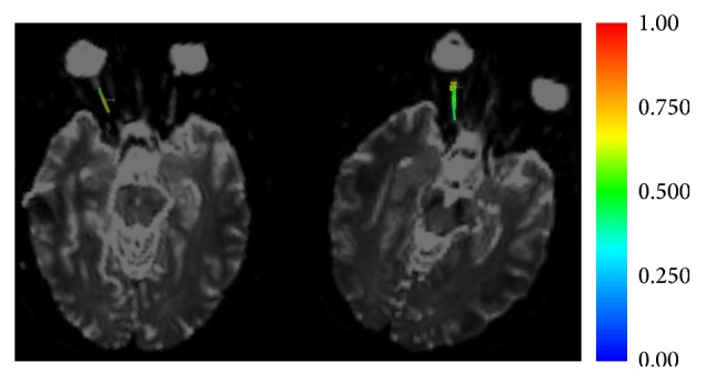
Glaucoma is a multifactorial disease that is the leading cause of irreversible blindness. Recent data documented that glaucoma is not limited to the retinal ganglion cells but that it also extends to the posterior visual pathway. The diagnosis is based on the presence of signs of glaucomatous optic neuropathy and consistent functional visual field alterations. Unfortunately these functional alterations often become evident when a significant amount of the nerve fibers that compose the optic nerve has been irreversibly lost. Advanced morphological and functional magnetic resonance (MR) techniques (morphometry, diffusion tensor imaging, arterial spin labeling, and functional connectivity) may provide a means for observing modifications induced by this fiber loss, within the optic nerve and the visual cortex, in an earlier stage. The aim of this systematic review was to determine if the use of these advanced MR techniques could offer the possibility of diagnosing glaucoma at an earlier stage than that currently possible.


Figures

Figure 1
Figure 1
Normal visual field examination (Humphrey field analyzer II 750 (Carl Zeiss Meditec, Inc., Dublin, CA; 30-2 test, full-threshold)) of a patient in an early stage of glaucoma.
Figure 2
Figure 2
Spectral domain optical coherence tomography (Cirrus, Carl Zeiss Meditec, Inc., Dublin, CA, version 6.5 software) of the same patient, showing the peripapillary RNFL (a) and macular GCL/IPL (ganglion cell layer/inner plexiform layer) (b) glaucomatous defects.
Figure 3
Figure 3
Cortical thickness estimates of the same patient as Figures 1 and 2. Note that the major concentration of the thinnest cortex (blue on the color scale) is located in the primary visual cortex and presents a greater extension on the left hemisphere.
Figure 4
Figure 4
DTI of the same patient as Figures 1 and 2. The fibers traced on the right optic nerve are colored with the fractional anisotropy measured at each point along the nerve (see legend). Note that the variation in color on the right (classic isometric sequence) is greater than that on the left (a priori sequence with the majority of the directions lying in the direction of the optic nerve). The greater variation indicates a greater variability in measurement: in other words the standard deviation of the sequence on the right is greater. The higher precision of the new sequence would be useful for the smaller modifications that should be present in the early stages of glaucoma.

References

    1. Casson R. J., Chidlow G., Wood J. P. M., Crowston J. G., Goldberg I. Definition of glaucoma: clinical and experimental concepts. Clinical and Experimental Ophthalmology. 2012;40(4):341–349. doi: 10.1111/j.1442-9071.2012.02773.x. - DOI - PubMed
    1. Harwerth R. S., Crawford M. L. J., Frishman L. J., Viswanathan S., Smith E. L., III, Carter-Dawson L. Visual field defects and neural losses from experimental glaucoma. Progress in Retinal and Eye Research. 2002;21(1):91–125. doi: 10.1016/s1350-9462(01)00022-2. - DOI - PubMed
    1. Rao H. L., Addepalli U. K., Chaudhary S., et al. Ability of different scanning protocols of spectral domain optical coherence tomography to diagnose preperimetric glaucoma. Investigative Ophthalmology and Visual Science. 2013;54(12):7252–7257. doi: 10.1167/iovs.13-12731. - DOI - PubMed
    1. Carpineto P., Ciancaglini M., Aharrh-Gnama A., Agnifili L., Mastropasqua L. Optical coherence tomography and retinal thickness analyzer features of spontaneous resolution of vitreomacular traction syndrome: a case report. European Journal of Ophthalmology. 2004;14(1):67–70. - PubMed
    1. Carpineto P., Nubile M., Agnifili L., et al. Reproducibility and repeatability of Cirrus HD-OCT peripapillary retinal nerve fibre layer thickness measurements in young normal subjects. Ophthalmologica. 2012;227(3):139–145. doi: 10.1159/000334967. - DOI - PubMed

Publication types

MeSH terms

LinkOut - more resources