. 2015:2015:203978.
doi: 10.1155/2015/203978. Epub 2015 Jun 18.

A Novel Matrine Derivative WM130 Inhibits Activation of Hepatic Stellate Cells and Attenuates Dimethylnitrosamine-Induced Liver Fibrosis in Rats

Affiliations

A Novel Matrine Derivative WM130 Inhibits Activation of Hepatic Stellate Cells and Attenuates Dimethylnitrosamine-Induced Liver Fibrosis in Rats

Yang Xu et al. Biomed Res Int. 2015.

Abstract

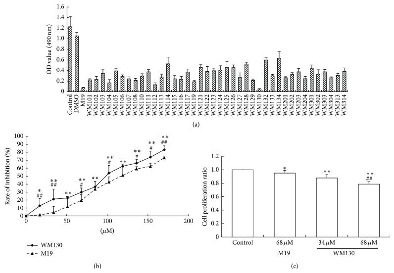
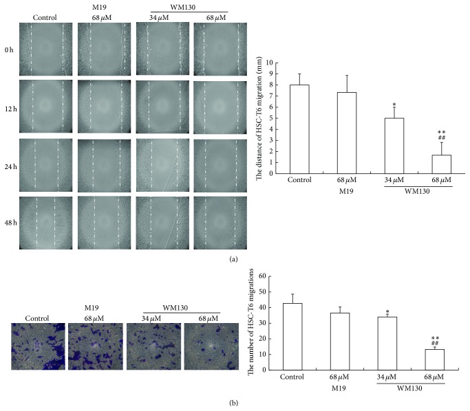
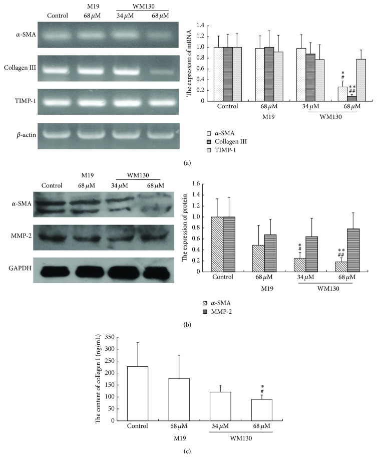
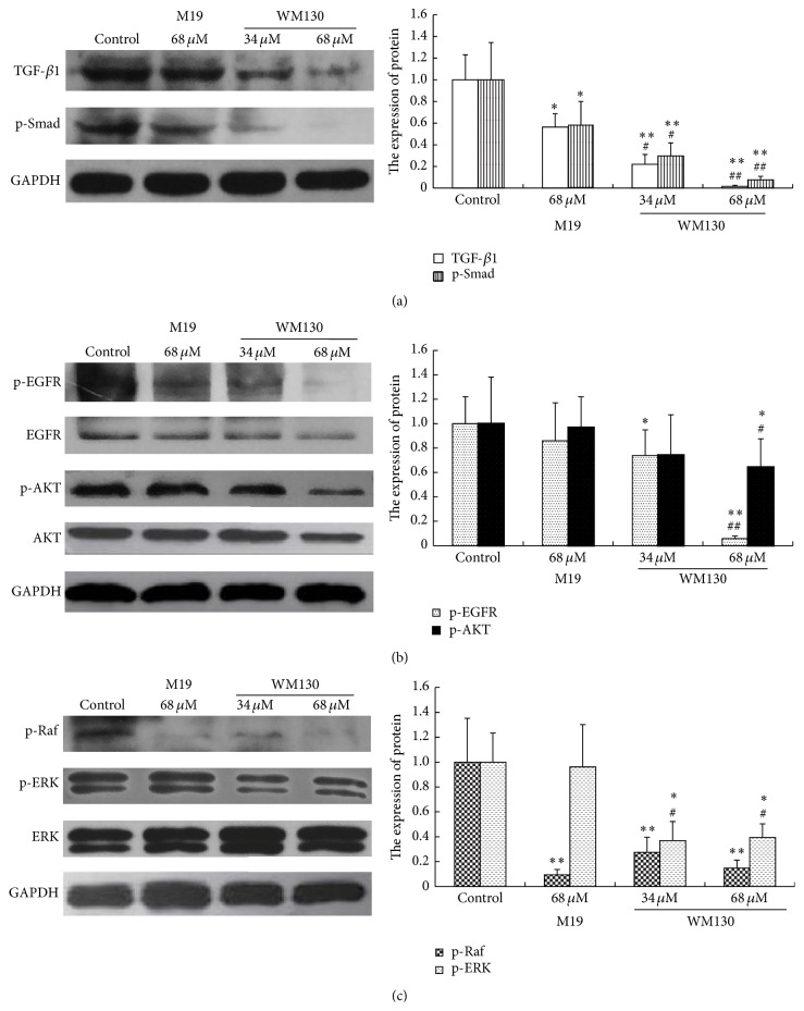
Activation of hepatic stellate cells (HSCs) is a critical event in process of hepatic fibrogenesis and cirrhosis. Matrine, the active ingredient of Sophora, had been used for clinical treatment of acute/chronic liver disease. However, its potency was low. We prepared a high potency and low toxicity matrine derivate, WM130 (C30N4H40SO5F), which exhibited better pharmacological activities on antihepatic fibrosis. This study demonstrated that WM130 results in a decreased proliferative activity of HSC-T6 cells, with the half inhibitory concentration (IC50) of 68 μM. WM130 can inhibit the migration and induce apoptosis in HSC-T6 cells at both concentrations of 68 μM (IC50) and 34 μM (half IC50). The expression of α-SMA, Collagen I, Collagen III, and TGF-β1 could be downregulated, and the protein phosphorylation levels of EGFR, AKT, ERK, Smad, and Raf (p-EGFR, p-AKT, p-ERK, p-Smad, and p-Raf) were also decreased by WM130. On the DMN-induced rat liver fibrosis model, WM130 can effectively reduce the TGF-β1, AKT, α-SMA, and p-ERK levels, decrease the extracellular matrix (ECM) formation, and inhibit rat liver fibrosis progression. In conclusion, this study demonstrated that WM130 can significantly inhibit the activation of HSC-T6 cells and block the rat liver fibrosis progression by inducing apoptosis, suppressing the deposition of ECM, and inhibiting TGF-β/Smad and Ras/ERK pathways.


Figures

Figure 1
Figure 1
Chemical structures of matrine and its derivatives, including M19 (C16N3H27S), and compound series. M19 was obtained through thiosulfate and side chain Michael addition, and the series of WM1, WM2, and WM3 were those containing an alkyl group or alkyl groups in place of one or more hydrogen atoms on M19. WM130 was obtained through reconstructing the structure of M19 with 18-methylamino-acylated and amino side chain addition.
Figure 2
Figure 2
WM130 and M19 inhibited the proliferation of HSC-T6 cells in vitro. (a) HSC-T6 cells were treated for 24 h with a series of different compounds at concentration of 0.1 mg/mL, and the cell viability was measured by MTT assay. (b) The effect of a series of different concentrations of WM130 or M19 was tested on cell viability of HSC-T6 cells. The inhibition rates were calculated by [inhibition rate = (1 − OD value of treatment group)/OD value of control group]. p < 0.05, ∗∗ p < 0.01 versus control group; # p < 0.05, ## p < 0.01 versus M19 group.
Figure 3
Figure 3
WM130 inhibited the migration and invasion of HSC-T6 cells. (a) The migration ability of HSC-T6 cells was analyzed by a scratch motility assay. WM130 68 μM group revealed a less migration distance during the treatment period of  0–48 h, compared with the control group or M19 group. (b) The effect of WM130 on inhibiting invasion of HSC-T6 cells was examined by transwell chamber assay. Invasive ability of HSC-T6 cells was quantified by counting the number of stained cells under microscopy (at 200x magnification). p < 0.05, ∗∗ p < 0.01 versus Control group; ## p < 0.01 versus M19 68 μM group.
Figure 4
Figure 4
WM130 induces apoptosis of HSC-T6 cells. (a) Blue stained nuclei were dyed by Hoechst and analyzed under fluorescent microscope (at 200x magnification). Apoptotic body could be observed in WM130 68 μM group (white arrow). More apoptotic HSC-T6 cells could be detected in WM130 68 μM group than in other treated group. (b) The Annexin V/PI apoptosis kit was used to quantify the percentage of cells undergoing apoptosis. The early apoptotic rates and the total apoptotic rates of HSC-T6 cells were detected in different groups by flow cytometry. ∗∗ p < 0.01 versus control group; # p < 0.05, ## p < 0.01 versus M19 68 μM group.
Figure 5
Figure 5
WM130 inhibits the activation of HSC-T6 cells and the secretion of ECM. (a) All samples were collected after incubation with WM130 and M19 for 24 h. Total RNA was extracted and the mRNA expression levels of α-SMA, Collagen I, and Collagen III in different groups were examined by RT-PCR assay. (b) Cells were lysed in RIPA lysis buffer supplemented with PMSF and the protein expression levels of α-SMA and MMP-2 in different groups were quantified by Western Blot assay. (c) Cell culture media in different groups were collected in which the concentrations of collagen I were measured by ELISA assay. All experiments were independently performed 3 times. p < 0.05, ∗∗ p < 0.01 versus control group; # p < 0.05, ## p < 0.01 versus M19 68 μM group.
Figure 6
Figure 6
WM130 inhibits TGF-β/Smad and Ras/ERK pathways. All samples were collected after incubation with WM130 and M19 for 24 h. Total protein was extracted by RIPA and the expression levels of (a) TGF-β1, p-Smad, (b) p-EGFR, p-AKT, and (c) p-Raf, p-ERK in HSC-T6 cells in different treated groups were determined by Western Blot assay. The experiments were independently performed 3 times. p < 0.05, ∗∗ p < 0.01 versus control group; # p < 0.05, ## p < 0.01 versus M19 68 μM group.
Figure 7
Figure 7
WM130 inhibits DMN-induced rat liver fibrosis. The model of liver fibrosis was induced by intraperitoneal injection with 1% DMN. Then rats were treated with M19 or WM130 by gavage. (a) The contents of the collagen fiber in liver tissue of DMN-induced rat were detected by Van Gieson's staining. The immunohistochemical staining was used to observe α-SMA, TGF-β1, p-AKT, and p-ERK positive areas in liver tissues of DMN-injured rats (at 200x magnification). (b) Sera were prepared from rat blood and used to determine the contents of ALT, AST, and Collagen I. ∗∗ p < 0.01, ∗∗∗ p < 0.001 versus normal Control; # p < 0.05, ### p < 0.001 versus model Control.

References

    1. Sakata K., Eda S., Lee E.-S., Hara M., Imoto M., Kojima S. Neovessel formation promotes liver fibrosis via providing latent transforming growth factor-β . Biochemical and Biophysical Research Communications. 2014;443(3):950–956. doi: 10.1016/j.bbrc.2013.12.074. - DOI - PubMed
    1. Hu L., Peng X., Tang Y., Liu Y. Synthesis of peptides of Carapax Trionycis and their inhibitory effects on TGF-β1-induced hepatic stellate cells. Drug Discoveries & Therapeutics. 2013;7(6):248–253. - PubMed
    1. Borg B. B., Seetharam A., Subramanian V., et al. Immune response to extracellular matrix collagen in chronic hepatitis C-induced liver fibrosis. Liver Transplantation. 2011;17(7):814–823. doi: 10.1002/lt.22303. - DOI - PMC - PubMed
    1. Dooley S., Delvoux B., Lahme B., Mangasser-Stephan K., Gressner A. M. Modulation of transforming growth factor β response and signaling during transdifferentiation of rat hepatic stellate cells to myofibroblasts. Hepatology. 2000;31(5):1094–1106. doi: 10.1053/he.2000.6126. - DOI - PubMed
    1. Nakao A., Afrakhte M., Morén A., et al. Identification of Smad7, a TGFβ-inducible antagonist of TGF-β signalling. Nature. 1997;389(6651):631–635. doi: 10.1038/39369. - DOI - PubMed

Publication types

MeSH terms