. 2015 Aug 15;69(5):509-18.
doi: 10.1097/QAI.0000000000000676.

Integrin α4β7 Expression Increases HIV Susceptibility in Activated Cervical CD4+ T Cells by an HIV Attachment-Independent Mechanism

Affiliations

Integrin α4β7 Expression Increases HIV Susceptibility in Activated Cervical CD4+ T Cells by an HIV Attachment-Independent Mechanism

Jian Ding et al. J Acquir Immune Defic Syndr. .

Abstract

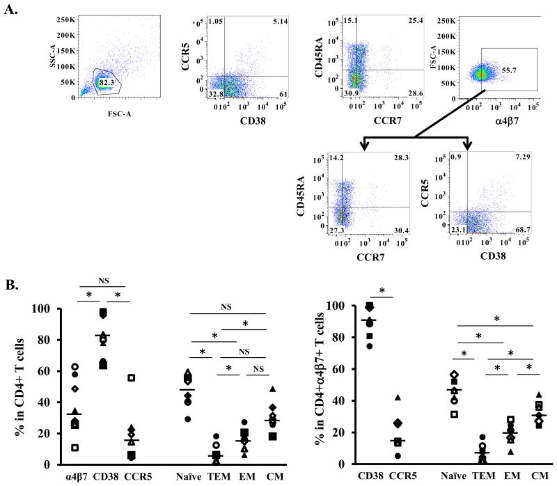
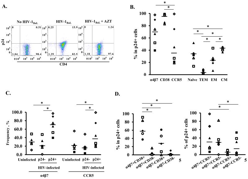
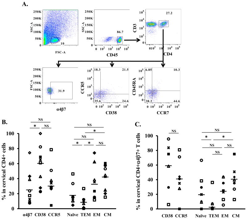
Background: CD4 T cells are crucial for the establishment and dissemination of HIV in mucosal tissues during acute infection. Studies indicate that integrin α4β7 CD4 T cells are preferentially infected by HIV in vitro and during acute SIV infection. The integrin α4β7 is thought to promote HIV capture by target cells; however, the role of integrin α4β7 in HIV transmission remains controversial. In this study, we characterized immune phenotypes of human cervical T cells and examined HIV preference in integrin α4β7 CD4 T cells. In vitro all-trans retinoic acid-differentiated peripheral CD4 T cells (atRA-differentiated cells) were included as a comparison.

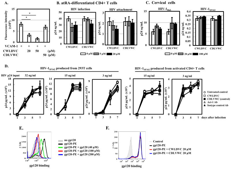
Results: In both peripheral and cervical cells, the majority of HIV p24 cells were activated CD4 T cells expressing integrin α4β7. Among infected atRA-differentiated cells, the frequency of CCR5 expression was higher in HIV p24 cells than in HIV p24 cells; no such difference was observed in cervical cells. Neither the cyclic hexapeptide CWLDVC nor a monoclonal antibody against integrin α4β7 blocked HIV attachment or gp120 binding to target cells regardless of the presence of CD4, indicating that integrin α4β7 did not facilitate HIV capture by target cells.

Conclusions: Integrin α4β7 expression increases HIV susceptibility, but the mechanism is not through promoting HIV binding to target cells.


Figures

Figure 1
Figure 1. Immunological phenotypes of atRA-differentiated CD4+ cells
Primary CD4+ T cells were isolated and then differentiated in the presence of atRA and anti-CD3 antibodies as described in Materials and Methods. (A) Gating strategy and analysis of cell surface expression of CD38, CCR5, CCR7, CD45RA, and integrin α4β7. T cell subsets were analyzed based on the following phenotypes: naïve (naïve cells, CD45RA+CCR7+), TEM (terminal effector memory cells, CD45RA+CCR7-), EM (effector memory cells, CD45RA-CCR7-) and CM (central memory cells, CD45RA-CCR7+). (B) Summary of percentages of cells expressing integrin α4β7, CD38 (activation marker), and CCR5 (HIV co-receptor) as well as different T cell subsets among total CD4+ population (left panel) and CD4+α4β7+ population (right panel) from 8 donors. Horizontal lines indicate the medians of the results from 8 donors. * indicates statistically significant difference (p ≤ 0.05, two-tailed Wilcoxon signed-rank test) between compared groups. NS, no significant difference (p>0.05).
Figure 2
Figure 2. Immunological phenotypes of atRA-differentiated CD4+ T cells that are preferentially infected by HIV
(A) atRA-differentiated CD4+ cells were infected by HIV-1BaL and HIV infection was monitored by FACS analysis with intracellular staining of HIV-1 Gag protein p24 at day 5 after infection. Cells were treated with the reverse transcriptase inhibitor AZT (10 μM) (right panel) to confirm the signal was from de novo synthesis of p24 rather than from HIV capture. (B) Percentage of cells expressing surface markers (integrin α4β7, CD38, CCR5) and of T cell subsets in HIV p24+ cells. (C) The frequency of cells expressing integrin α4β7 or CCR5 in HIV p24+ and HIVp24- populations in HIV-infected cells. Uninfected cells are included as a comparison. * P<0.05, two-tailed Wilcoxon signed-rank test. (D) Among HIV p24+ T cells, summary of analysis of co-expression of α4β7 and CD38 (left panel) and of α4β7 and CCR5 (right panel) from 6 donors. *p<0.05, two-tailed Wilcoxon signed-rank test.
Figure 3
Figure 3. Immunological phenotypes of cervical CD4+ T cells
Single-cell suspensions from cervical tissues without gross pathology from women undergoing therapeutic hysterectomy were prepared as described in materials and methods. (A) Gating strategy and FACS analysis of cell surface markers in samples from one donor. The lymphocyte populations were identified according to their light-scattering properties. Leukocytes were further identified by the expression of CD45. CD4+ T cells among leukocytes were gated as CD3+CD4+ cells. Expression of integrin α4β7, CD38, CCR5, CCR7, and CD45RA on the cervical CD4+ T cells was then analyzed. (B) Summary of percentage of CD4+ cervical cells expressing specific markers and T cell subsets from 9 donors. (C). Frequencies of cervical CD4+α4β7+ T cells expressing specific markers and T cell subsets from 9 donors. * p<0.05, statistically significant difference, two-tailed Wilcoxon signed-rank test. NS, no significant difference (p>0.05).
Figure 4
Figure 4. Immunological phenotypes of cervical CD4+ T cells that are preferentially infected by HIV
Cervical cells were exposed to HIV-1BaL for 2 h. After washing off unbound virus, infected cells were cultured for 5 days before FACS analysis. (A) Summary of HIV preference in cervical CD4+ T cells from 7 donors. The p24+ cells were back-gated onto various cell markers including integrin α4β7, CD38, CCR5, and CD45RA. (B) Frequencies of cells expressing integrin α4β7, CD38, or CCR5 in HIV p24+ and HIVp24- populations in HIV-infected cells. Uninfected cells are included as a comparison. *p<0.05, statistically significant difference, two-tailed Wilcoxon-sign rank test; NS, not significant. (C) Frequencies of HIV p24+CD4+ T cells that were co-expressed α4β7 and CD38 (left panel) or of α4β7 and CCCR5 (right panel).
Figure 5
Figure 5. Integrin α4β7 does not promote HIV binding to atRA-differentiated CD4+ T cells or cervical cells
(A) Effect of integrin α4β7 binding peptide CWLDVC or control peptide (CDLVWC) on VCAM-1 mediated cell adhesion was determined as described in materials and methods. (B) atRA-differentiated CD4+ cells or (C) cervical cells were pretreated with integrin α4β7 binding peptide or control peptide at the indicated concentrations for 1 h at 37°C prior to HIV exposure for 2 h. For HIV infection, peptides were added back after washing off unbound virus and viral production in the culture media was determined by p24 ELISA at day 7 after infection. For HIV attachment, cells were exposed to HIV for 2 h at 4°C in the presence of peptides. After washing with ice-cold PBS, the cells were pelleted and lysed in PBS with 1% Triton X-100. Cell-associated HIV was determined by p24 ELISA. (D) HIV-1SF162 was produced from either 293T cells by transfection or from PHA-activated CD4+ T cells. PHA-activated CD4+ T cells (1×10⁵ cells per well in a 96-well plate) were pre-treated with integrin α4β7 binding peptide (100 μM), control peptide (100 μM), Act-1 Ab (5 μg/ml) or isotype control Ab (5 μg/ml) for 1 h before exposure to different concentrations of HIV-1SF162 from 293T cells or activated CD4+T cells. After 2 h incubation, cells were washed and peptides or antibodies were added back. HIV replication was monitored at different time points by measuring HIV p24 in media using HIVp24 alphaLISA kits. (E) Binding specificity of HIV gp120 to PHA-activated PBLs. Cells were incubated with gp120-PE (20 μM) at 4°C for 1 h in the presence of unconjugated gp120 at indicated concentrations followed by FACS analysis. (F) Effect of integrin α4β7 binding peptide on HIV gp120 binding to CD4+ T cells. PHA-activated PBLs were incubated with the CWLDVC peptide or the control peptide (CDLVWC) for 1 h at 4°C before addition of HIV gp120-PE at 20 μM in the presence of peptides and bivalent ions (100 mM CaCl2 and 1 mM MnCl2 in PBS) for an additional hour. HIV gp120 biding to target cells was then assessed by FACS analysis. *p<0.05, statistically significant difference, two-tailed Student's t test. Results were representatives of three independent experiments from different donors.

References

    1. Haase AT. Early events in sexual transmission of HIV and SIV and opportunities for interventions. Annu Rev Med. 2011 Feb 18;62:127–39. - PubMed
    1. Cicala C, Martinelli E, McNally JP, et al. The integrin alpha4beta7 forms a complex with cell-surface CD4 and defines a T-cell subset that is highly susceptible to infection by HIV-1. Proc Natl Acad Sci U S A. 2009 Dec 8;106(49):20877–82. - PMC - PubMed
    1. Kader M, Bixler S, Roederer M, Veazey R, Mattapallil JJ. CD4 T cell subsets in the mucosa are CD28+Ki-67-HLA-DR-CD69+ but show differential infection based on alpha4beta7 receptor expression during acute SIV infection. J Med Primatol. 2009 Oct;38(Suppl 1):24–31. - PMC - PubMed
    1. Kader M, Wang X, Piatak M, et al. Alpha4(+)beta7(hi)CD4(+) memory T cells harbor most Th-17 cells and are preferentially infected during acute SIV infection. Mucosal Immunol. 2009 Sep;2(5):439–49. - PMC - PubMed
    1. Arthos J, Cicala C, Martinelli E, et al. HIV-1 envelope protein binds to and signals through integrin alpha4beta7, the gut mucosal homing receptor for peripheral T cells. Nat Immunol. 2008 Mar;9(3):301–9. - PubMed

Publication types