. 2015 Aug;22(8):618-26.
doi: 10.1038/nsmb.3055. Epub 2015 Jul 13.

A unique binding mode enables MCM2 to chaperone histones H3-H4 at replication forks

Affiliations

A unique binding mode enables MCM2 to chaperone histones H3-H4 at replication forks

Hongda Huang et al. Nat Struct Mol Biol. 2015 Aug.

Abstract

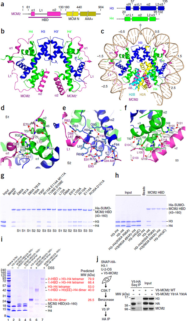
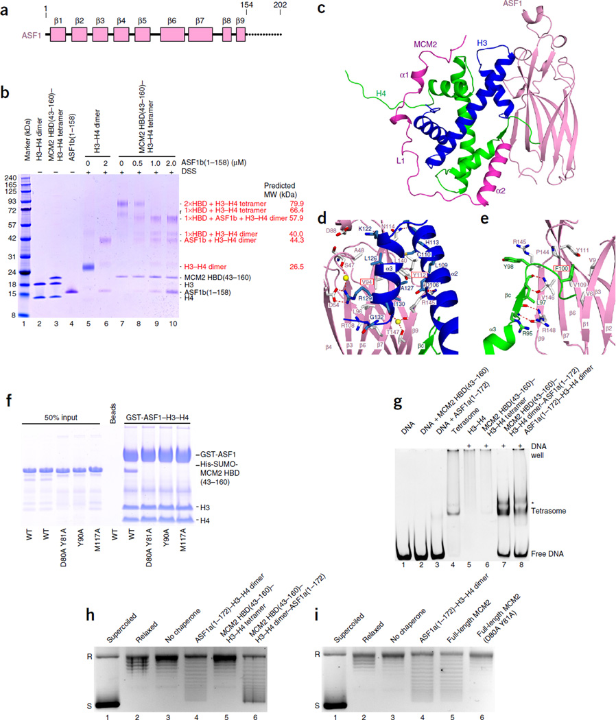
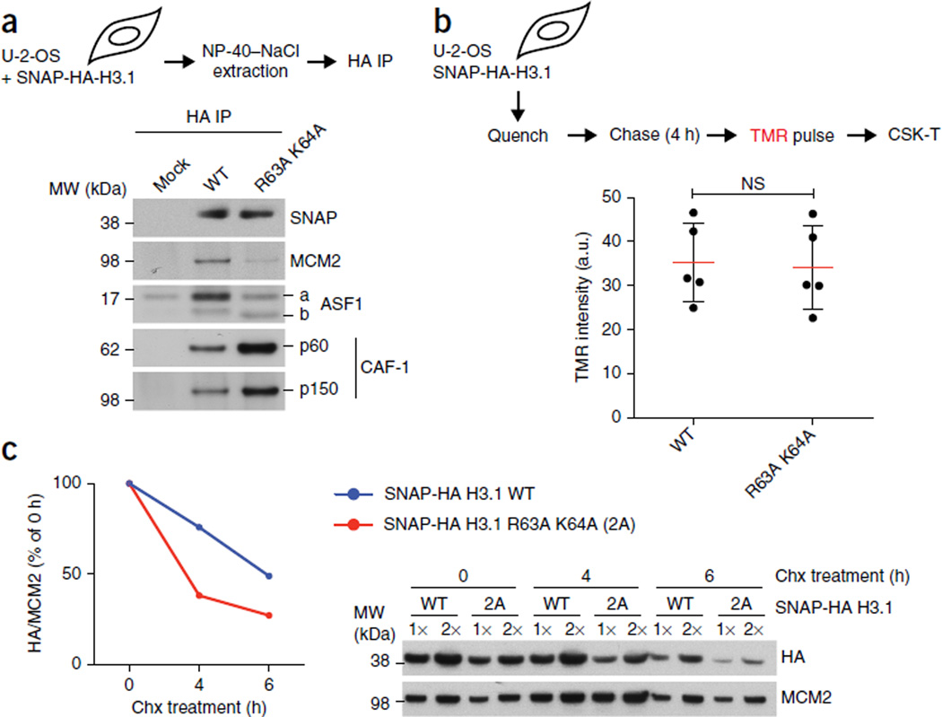
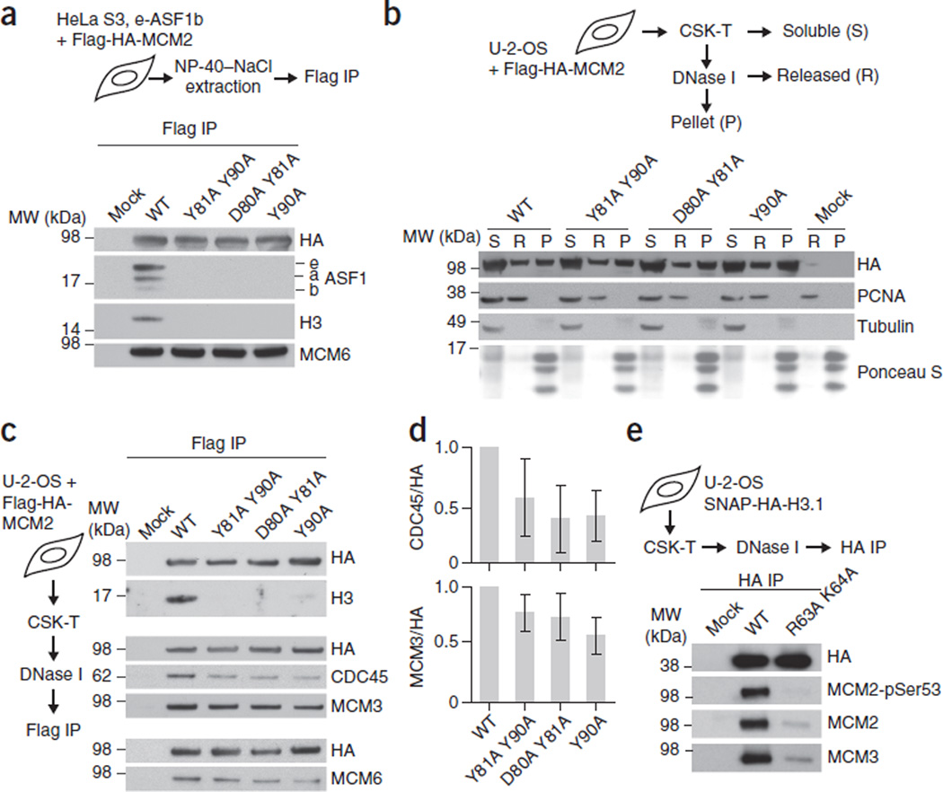
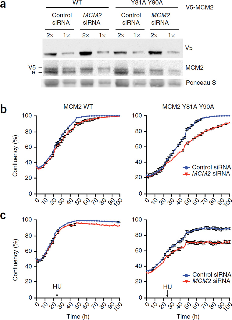
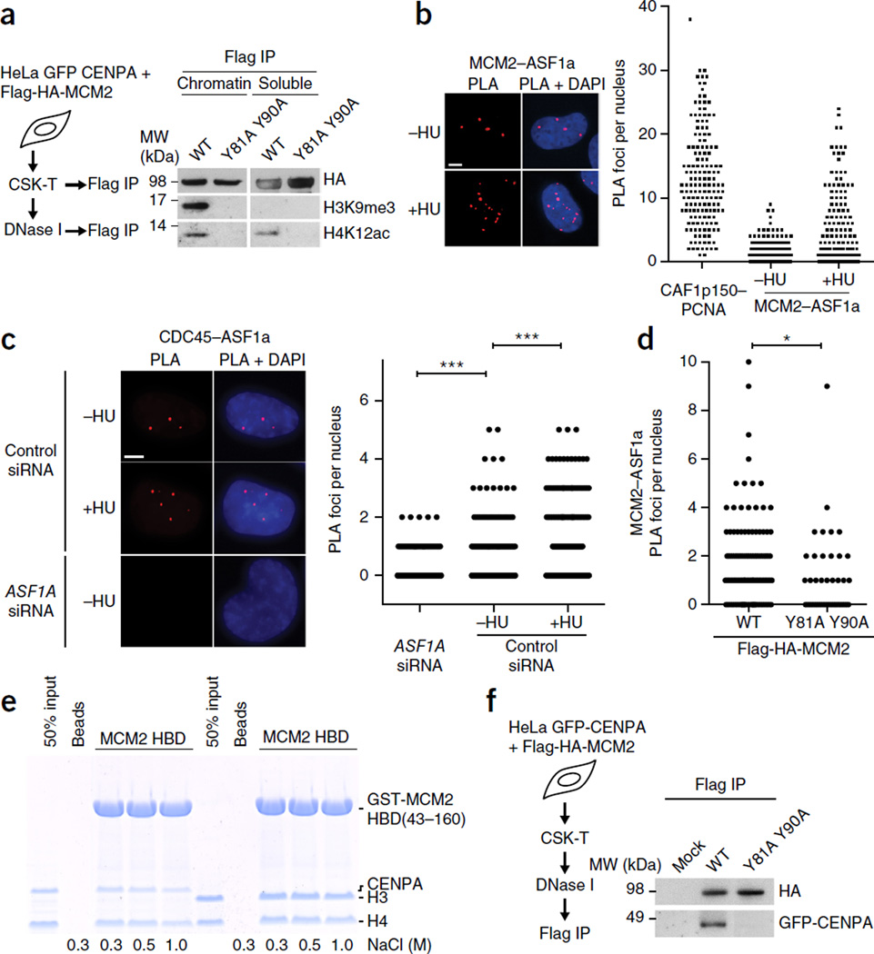
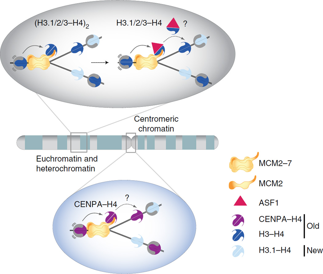
During DNA replication, chromatin is reassembled by recycling of modified old histones and deposition of new ones. How histone dynamics integrates with DNA replication to maintain genome and epigenome information remains unclear. Here, we reveal how human MCM2, part of the replicative helicase, chaperones histones H3-H4. Our first structure shows an H3-H4 tetramer bound by two MCM2 histone-binding domains (HBDs), which hijack interaction sites used by nucleosomal DNA. Our second structure reveals MCM2 and ASF1 cochaperoning an H3-H4 dimer. Mutational analyses show that the MCM2 HBD is required for MCM2-7 histone-chaperone function and normal cell proliferation. Further, we show that MCM2 can chaperone both new and old canonical histones H3-H4 as well as H3.3 and CENPA variants. The unique histone-binding mode of MCM2 thus endows the replicative helicase with ideal properties for recycling histones genome wide during DNA replication.


Figures

Figure 1
Figure 1
Structure of the MCM2 HBD–H3–H4 tetramer complex. (a) Schematics of domain architectures of MCM2, H3 and H4. (b) Ribbon representation of the structure of the MCM2 HBD–H3–H4 tetramer complex. (c) A model involving replacement of the H3–H4 tetramer in the nucleosome structure (PDB 1AOI) by the structure of the MCM2 HBD–H3–H4 tetramer complex. (d–f) Details of the interactions between the MCM2 HBD and an H3–H4 tetramer spanning regions S1 (d), S2 (e) and S3 (f) of the complex. (g) Pulldowns of HBD mutants with H3–H4 tetramers. (h) Pulldowns of HBD WT with H3 and H4 mutants. (i) DSS cross-linking under physiological salt (150 mM). Lanes 2–4, controls without DSS; lane 5, H3–H4 cross-linked as a dimer; lane 6, cross-linked MCM2(43–160)–H3(EE)–H4 dimer, with H3(EE) denoting the L126E I130E mutant; lane 7, cross-linked MCM2(43–160)–H3–H4 tetramer. (j) Sequential (seq) IP of V5-MCM2 and SNAP-HA-H3.1 from solubilized chromatin. U-2-OS cells stably expressing SNAP-HA-H3.1 were transfected with V5-MCM2 WT or Y81A Y90A mutant as indicated. The V5-MCM2 complexes were isolated and then eluted with V5 peptide, which were subjected to a second IP with anti-HA before western blot analysis. Uncropped images of gels are shown in Supplementary Data Set 1. MW, molecular weight; His, histidine tag.
Figure 2
Figure 2
Structure of the MCM2 HBD–H3–H4 dimer–ASF1 complex. (a) Schematics of domain architecture of ASF1. (b) DSS cross-linking results showing the transition of MCM2-bound H3–H4 tetramer into dimer by addition of ASF1 under physiological salt (150 mM). Lanes 2–4, controls without DSS; lanes 5–10, titration of ASF1 into H3–H4 dimer or MCM2 HBD(43–160)–H3–H4 tetramer complex, with cross-linking after titrations. (c) Ribbon representation of the structure of the MCM2 HBD–H3–H4 dimer–ASF1 complex. (d,e) Details of the intermolecular interactions of ASF1 with H3 (d) and H4 (e) in the MCM2 HBD–H3–H4 dimer–ASF1 complex. (f) Pulldowns of immobilized GST-ASF1–H3–H4 dimer complex with key mutants of MCM2 HBD. (g) Tetrasome assembly on linear DNA, monitored by native PAGE. Lanes 1–4, controls; lanes 5–8, H3–H4 tetramer or different prepurified chaperone–H3–H4 complexes incubated with linear DNA. Asterisk, unassigned band. (h,i) Plasmid-supercoiling assays showing chaperone activities of the prepurified MCM2 HBD–H3–H4 dimer–ASF1 complex (h) and the full-length MCM2 (i). R, relaxed DNA; S, supercoiled DNA. Uncropped images of gels are shown in Supplementary Data Set 1.
Figure 3
Figure 3
MCM2 binding stabilizes non-nucleosomal H3.1–H4. (a) IP of SNAP-HA-H3.1 WT or R63A K64A mutant from transfected cells (input material for IP in Supplementary Fig. 5a). (b) Replication-coupled H3.1 incorporation, measured by SNAP-tag fluorescent tetramethylrhodamine (TMR) labeling in stable cells expressing SNAP-HA-H3.1 WT or R63A K64A. Top, schematic showing quenching of old SNAP-HA-H3.1 to block labeling; incorporation of newly synthesized histones for 4 h before TMR labeling; and preextraction with CSK 0.5% Triton X-100 (CSK-T). Bottom, dot plot showing mean (red lines) TMR intensities are from five independent cell cultures and experiments, each including five technical replicates including more than 5,000 TMR-positive cells (representative micrographs in Supplementary Fig. 6a). Error bars (black lines), s.d.; NS, not significant by two-tailed unpaired t test; a.u., arbitrary units. (c) Stability of non-nucleosomal H3.1 WT and R63A K64A. Left, quantification, with SNAP-HA-H3.1 levels shown relative to MCM2. Right, western blot. Samples are soluble histones extracted from stable SNAP-HA-H3.1 WT or mutant cell lines synchronized in S phase and treated with hydroxyurea (HU) and cycloheximide (chx) to inhibit replication-coupled histone deposition and protein synthesis, respectively. One representative experiment out of three is shown. Uncropped images of gels are shown in Supplementary Data Set 1.
Figure 4
Figure 4
MCM2 chaperones H3–H4 as part of the MCM2–7 helicase in chromatin. (a) IP of transiently expressed Flag-HA-MCM2 from extracts of HeLa S3 cells stably expressing e-ASF1b. MCM2 mutations were designed to disrupt interactions with H3 (Y90A), H4 (D80A Y81A) and both H3 and H4 (Y81A Y90A). (b) Fractionation of cells expressing Flag-HA-MCM2 mutants. α-tubulin, fractionation control. (c) IP of Flag-HA-MCM2 mutants from solubilized chromatin. (d) Quantification of coimmunoprecipitated CDC45 and MCM3 relative to Flag-HA-MCM2. Error bars, s.d. from the mean for Y81A Y90A (n = 4 independent cell cultures and experiments), D80A Y81A (n = 4) and Y90A (n = 3). (e) IP of SNAP-HA-H3.1 WT or mutant from DNase I–solubilized chromatin of stable cell lines (input material for IPs in Supplementary Fig. 5b–d). pSer53, phosphorylated Ser53. Uncropped images of gels are shown in Supplementary Data Set 1.
Figure 5
Figure 5
MCM2 histone-chaperone function is required for cell proliferation. (a–c) U-2-OS cells transduced with siRNA-resistant MCM2 WT or Y81A Y90A, treated with MCM2 siRNA and analyzed by western blotting (e, endogenous) (a) or high-content live-cell imaging to detect cell proliferation in the absence (b) or presence (c) of HU. Imaging was started 6 h after siRNA transfection; HU (0.5 mM) was added 24 h later as indicated. The graphs show the mean confluency (%) ± range from n = 2 technical replicates and are representative of three independent experiments and cell cultures. Uncropped images of gels are shown in Supplementary Data Set 1.
Figure 6
Figure 6
MCM2 can handle new and old histones, including all H3 variants. (a) IP of Flag-HA-MCM2 WT and mutant from soluble fractions or DNase I–solubilized chromatin. (b,c) PLA of endogenous chromatin-bound ASF1a with MCM2 (b) or CDC45 (c). Soluble proteins were extracted by CSK-T. Positive control, PLA of CAF-1 p150 and PCNA; negative control, PLA in cells depleted for ASF1a. Dot plots show PLA interaction foci per nucleus and are representative of two independent cell cultures and stainings. In c, n = 270 cells (ASF1A siRNA), n = 260 cells (control siRNA, −HU) and n = 284 cells (control siRNA, +HU). ***P < 0.001 by unpaired two-tailed t test with Welch’s correction. (d) PLA of endogenous chromatin-bound ASF1a and MCM2 in U-2-OS cells transiently transfected with Flag-HA-MCM2 WT or Y81A Y90A mutant. Dot plot shows PLA interaction foci per nucleus and is representative of two independent cell cultures and stainings. n = 188 (WT); n = 44 (Y81A Y90A). *P < 0.05 by unpaired two-tailed t test with Welch’s correction. (Representative images are shown in Supplementary Fig. 7c.) (e) Pulldowns of GST-MCM2 HBD with H3–H4 or CENPA–H4 tetramers in different salt conditions. (f) IP of Flag-HA-MCM2 WT and mutant from DNase I–solubilized chromatin of cells stably expressing GFP-CENPA. (Input material for IP is shown in Supplementary Fig. 5f. GFP-CENPA coprecipitation with MCM2 in CSK-T fractions is shown in Supplementary Fig. 8d.) Uncropped images of gels are shown in Supplementary Data Set 1.
Figure 7
Figure 7
Structure-based model for how MCM2 handles parental histones H3 and H4 genome wide during DNA replication. H3–H4 or CENPA–H4 tetramers, released from parental nucleosomes upon progression of the MCM2–7 helicase, are captured by MCM2 and held in proximity to the fork. MCM2-bound H3.1–H4, H3.2–H4 or H3.3–H4 tetramers can be disrupted by ASF1, which in turn could mediate redeposition of the dimers. Alternatively, other chaperones such as FACT might mediate histone transfer. Chaperone choice could be context dependent; for example, a CENPA–H4–specific chaperone should be brought in during replication of centromeric DNA. A key feature of this model is the ability of MCM2 to hijack the nucleosomal DNA-binding surface to chaperone H3–H4 tetramers, thus shielding the histones and ensuring their recycling regardless of their subtype and modifications. H3.1/2/3, histone H3 variants H3.1, H3.2 and H3.3.

Comment in

References

    1. Alabert C, Groth A. Chromatin replication and epigenome maintenance. Nat. Rev. Mol. Cell Biol. 2012;13:153–167. - PubMed
    1. Hake SB, Allis CD. Histone H3 variants and their potential role in indexing mammalian genomes: the “H3 barcode hypothesis”. Proc. Natl. Acad. Sci. USA. 2006;103:6428–6435. - PMC - PubMed
    1. Margueron R, Reinberg D. Chromatin structure and the inheritance of epigenetic information. Nat. Rev. Genet. 2010;11:285–296. - PMC - PubMed
    1. Probst AV, Dunleavy E, Almouzni G. Epigenetic inheritance during the cell cycle. Nat. Rev. Mol. Cell Biol. 2009;10:192–206. - PubMed
    1. Shibahara K, Stillman B. Replication-dependent marking of DNA by PCNA facilitates CAF-1-coupled inheritance of chromatin. Cell. 1999;96:575–585. - PubMed

Publication types

MeSH terms