

Quantitative Evaluation of Rabbit Brain Injury after Cerebral Hemisphere Radiation Exposure Using Generalized q-Sampling Imaging
- PMID: 26168047
- PMCID: PMC4500591
- DOI: 10.1371/journal.pone.0133001
Quantitative Evaluation of Rabbit Brain Injury after Cerebral Hemisphere Radiation Exposure Using Generalized q-Sampling Imaging
Abstract
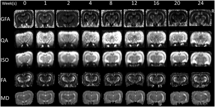
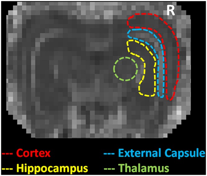
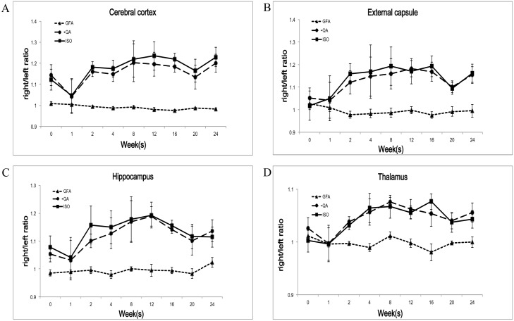
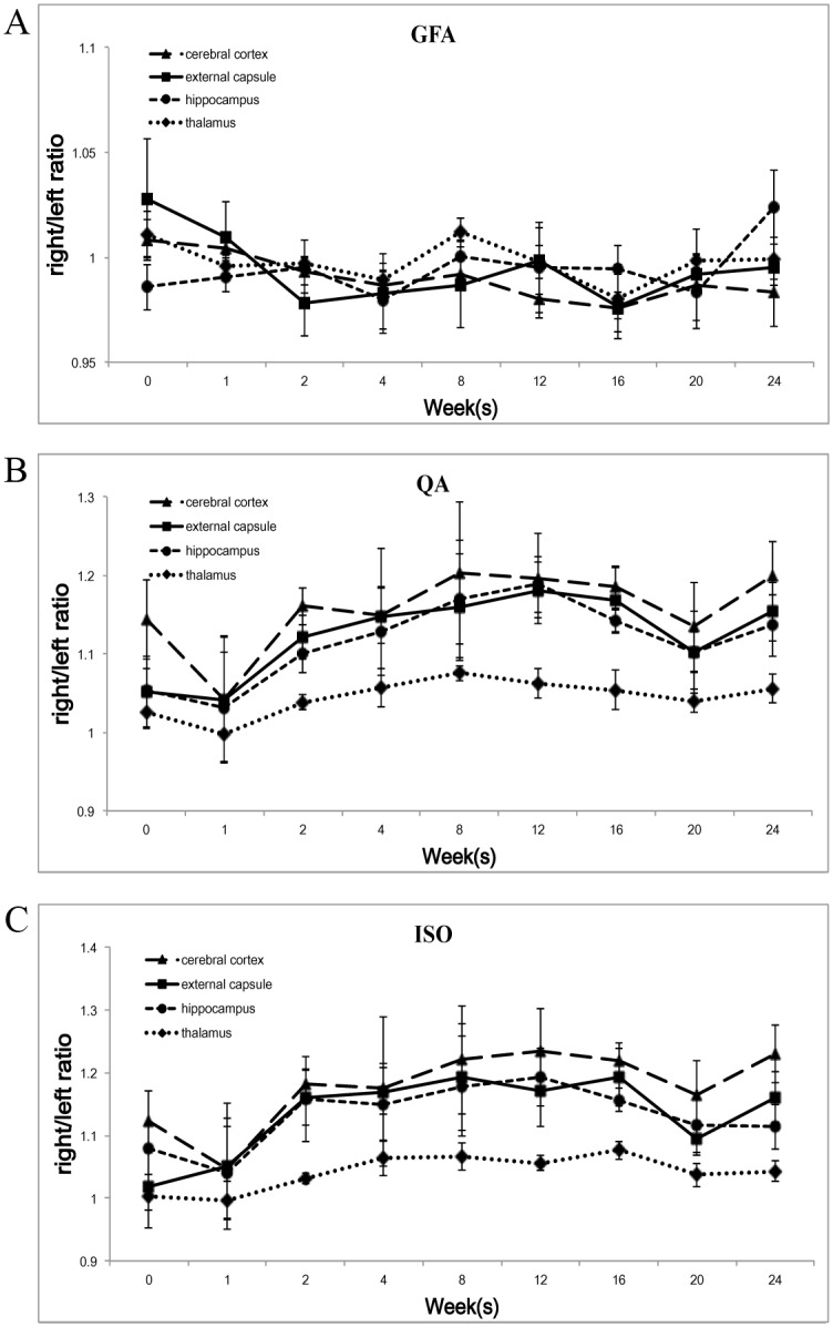
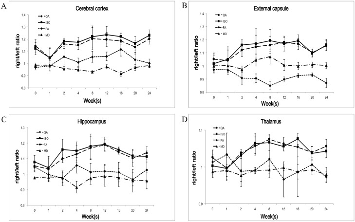
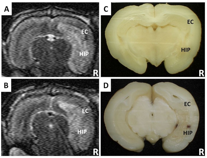
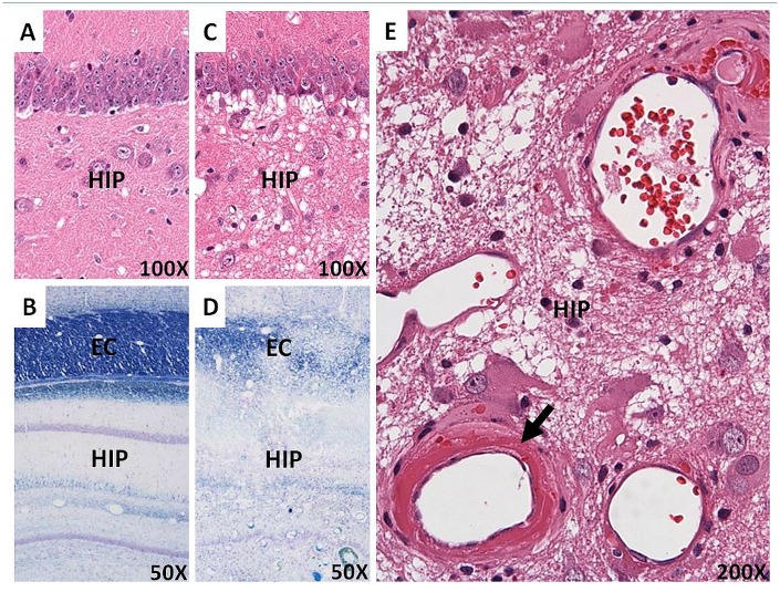
Radiation therapy is widely used for the treatment of brain tumors and may result in cellular, vascular and axonal injury and further behavioral deficits. The non-invasive longitudinal imaging assessment of brain injury caused by radiation therapy is important for determining patient prognoses. Several rodent studies have been performed using magnetic resonance imaging (MRI), but further studies in rabbits and large mammals with advanced magnetic resonance (MR) techniques are needed. Previously, we used diffusion tensor imaging (DTI) to evaluate radiation-induced rabbit brain injury. However, DTI is unable to resolve the complicated neural structure changes that are frequently observed during brain injury after radiation exposure. Generalized q-sampling imaging (GQI) is a more accurate and sophisticated diffusion MR approach that can extract additional information about the altered diffusion environments. Therefore, herein, a longitudinal study was performed that used GQI indices, including generalized fractional anisotropy (GFA), quantitative anisotropy (QA), and the isotropic value (ISO) of the orientation distribution function and DTI indices, including fractional anisotropy (FA) and mean diffusivity (MD) over a period of approximately half a year to observe long-term, radiation-induced changes in the different brain compartments of a rabbit model after a hemi-brain single dose (30 Gy) radiation exposure. We revealed that in the external capsule, the GFA right to left (R/L) ratio showed similar trends as the FA R/L ratio, but no clear trends in the remaining three brain compartments. Both the QA and ISO R/L ratios showed similar trends in the all four different compartments during the acute to early delayed post-irradiation phase, which could be explained and reflected the histopathological changes of the complicated dynamic interactions among astrogliosis, demyelination and vasogenic edema. We suggest that GQI is a promising non-invasive technique and as compared with DTI, it has better potential ability in detecting and monitoring the pathophysiological cascades in acute to early delayed radiation-induced brain injury by using clinical MR scanners.
Conflict of interest statement
Figures
Similar articles
-
New insights into the developing rabbit brain using diffusion tensor tractography and generalized q-sampling MRI.PLoS One. 2015 Mar 23;10(3):e0119932. doi: 10.1371/journal.pone.0119932. eCollection 2015. PLoS One. 2015. PMID: 25798595 Free PMC article.
-
Diffusion tensor imaging during recovery from severe traumatic brain injury and relation to clinical outcome: a longitudinal study.Brain. 2008 Feb;131(Pt 2):559-72. doi: 10.1093/brain/awm294. Epub 2007 Dec 14. Brain. 2008. PMID: 18083753
-
Characterizing longitudinal changes in rabbit brains infected with Angiostrongylus Cantonensis based on diffusion anisotropy.Acta Trop. 2016 May;157:1-11. doi: 10.1016/j.actatropica.2016.01.020. Epub 2016 Jan 22. Acta Trop. 2016. PMID: 26808581
-
The role of diffusion tensor imaging in the evaluation of ischemic brain injury - a review.NMR Biomed. 2002 Nov-Dec;15(7-8):561-9. doi: 10.1002/nbm.786. NMR Biomed. 2002. PMID: 12489102 Review.
-
Diffusion-weighted MR of the brain: methodology and clinical application.Radiol Med. 2005 Mar;109(3):155-97. Radiol Med. 2005. PMID: 15775887 Review. English, Italian.
Cited by
-
Microstructural Properties of Brain White Matter Tracts in Breast Cancer Survivors: A Diffusion Tensor Imaging Study.Pathophysiology. 2022 Oct 17;29(4):595-609. doi: 10.3390/pathophysiology29040046. Pathophysiology. 2022. PMID: 36278563 Free PMC article.
-
Tay-Sachs and Sandhoff Diseases: Diffusion tensor imaging and correlational fiber tractography findings differentiate late-onset GM2 Gangliosidosis.medRxiv [Preprint]. 2024 Dec 16:2024.12.13.24318793. doi: 10.1101/2024.12.13.24318793. medRxiv. 2024. Update in: J Neurol. 2025 Apr 23;272(5):355. doi: 10.1007/s00415-025-13091-3. PMID: 39802759 Free PMC article. Updated. Preprint.
-
Evaluation of structural connectivity changes in betel-quid chewers using generalized q-sampling MRI.Psychopharmacology (Berl). 2017 Jul;234(13):1945-1955. doi: 10.1007/s00213-017-4602-0. Epub 2017 Mar 24. Psychopharmacology (Berl). 2017. PMID: 28342092
-
White Matter Microstructural Alterations in Newly Diagnosed Parkinson's Disease: A Whole-Brain Analysis Using dMRI.Brain Sci. 2022 Feb 7;12(2):227. doi: 10.3390/brainsci12020227. Brain Sci. 2022. PMID: 35203990 Free PMC article.
-
Gender differences in the structural connectome of the teenage brain revealed by generalized q-sampling MRI.Neuroimage Clin. 2017 May 22;15:376-382. doi: 10.1016/j.nicl.2017.05.014. eCollection 2017. Neuroimage Clin. 2017. PMID: 28580294 Free PMC article.
References
-
- Stone HB, Coleman CN, Anscher MS, McBride WH. Effects of radiation on normal tissue: consequences and mechanisms. Lancet Oncol. 2003;4(9):529–36. Epub 2003/09/11. doi: S1470204503011914 [pii]. . - PubMed
-
- Tofilon PJ, Fike JR. The radioresponse of the central nervous system: a dynamic process. Radiat Res. 2000;153(4):357–70. Epub 2000/05/08. . - PubMed
Publication types
MeSH terms
LinkOut - more resources
Full Text Sources
Other Literature Sources
