

Protective Macroautophagy Is Involved in Vitamin E Succinate Effects on Human Gastric Carcinoma Cell Line SGC-7901 by Inhibiting mTOR Axis Phosphorylation
- PMID: 26168048
- PMCID: PMC4500415
- DOI: 10.1371/journal.pone.0132829
Protective Macroautophagy Is Involved in Vitamin E Succinate Effects on Human Gastric Carcinoma Cell Line SGC-7901 by Inhibiting mTOR Axis Phosphorylation
Abstract
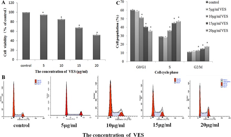
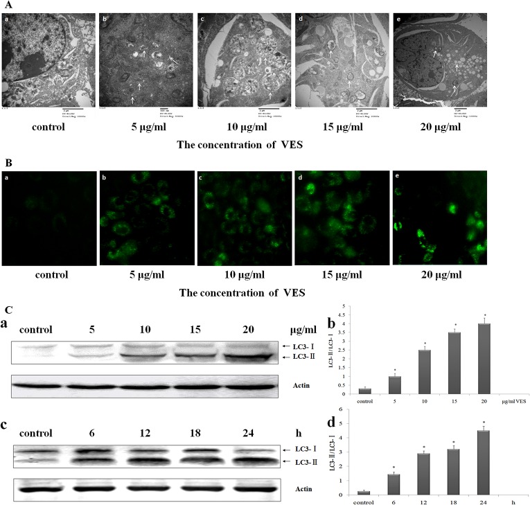
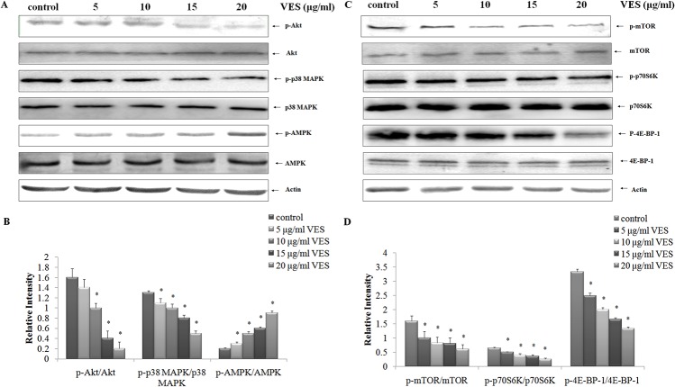
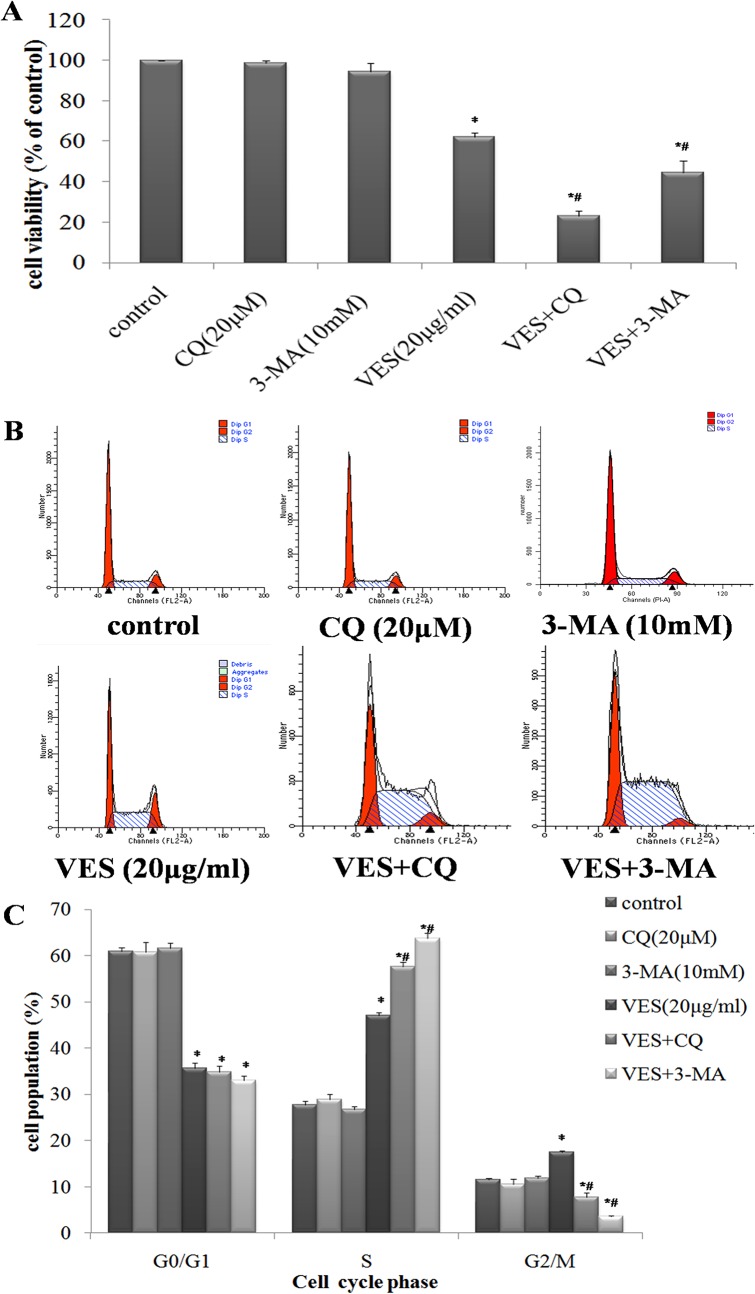
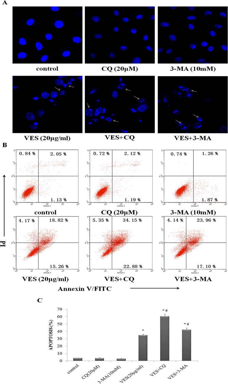
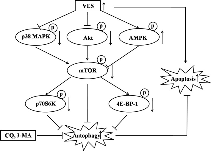
Vitamin E succinate (VES), a potential cancer therapeutic agent, potently induces apoptosis and inhibits the growth of various cancer cells. Autophagy has been supposed to promote cancer cell survival or trigger cell death, depending on particular cancer types and tumor microenvironments. The role of autophagy in the growth suppressive effect of VES on gastric cancer cell is basically unknown. We aimed to determine whether and how autophagy affected the VES-induced inhibition of SGC-7901 human gastric carcinoma cell growth. SGC-7901 cells were treated with VES or pre-treated with autophagy inhibitor, chloroquine (CQ) and 3-methyladenine (3-MA). Electron microscopy, fluorescence microscopy and Western blot were used to study whether VES induced autophagy reaction in SGC-7901 cells. Western blot evaluated the activities of the mammalian target of rapamycin (mTOR) axis. Then we used 3-(4,5-dimethylthiazol-2-yl)-2,5-diphenyltetrazolium bromide (MTT) and flow cytometry to detect the level of cell viability and apoptosis. Collectively, our data indeed strongly support our hypothesis that VES treatment produced cytological variations that depict autophagy, increased the amount of intracellular green fluorescent protein-microtubule associated protein 1 light chain 3 (GFP-LC3) punctate fluorescence and the number of autophagic vacuoles. It altered the expression of endogenous autophagy marker LC3. VES activated the suppression of mTOR through inhibiting upstream regulators p38 MAPK and Akt. mTOR suppression consequently inhibited the activation of mTOR downstream targets p70S6K and 4E-BP-1. The activation of the upstream mTOR inhibitor AMPK had been up-regulated by VES. The results showed that pre-treatment SGC-7901 with autophagy inhibitors before VES treatment could increase the capacity of VES to reduce cell viability and to provoke apoptosis. In conclusion, VES-induced autophagy participates in SGC-7901 cell protection by inhibiting mTOR axis phosphorylation. Our findings not only strengthen our understanding of the roles of autophagy in cancer biology, but may also be useful for developing new treatments for gastric cancer patients.
Conflict of interest statement
Figures
Similar articles
-
Akt/AMPK/mTOR pathway was involved in the autophagy induced by vitamin E succinate in human gastric cancer SGC-7901 cells.Mol Cell Biochem. 2017 Jan;424(1-2):173-183. doi: 10.1007/s11010-016-2853-4. Epub 2016 Oct 28. Mol Cell Biochem. 2017. PMID: 27796683
-
E Platinum, a newly synthesized platinum compound, induces autophagy via inhibiting phosphorylation of mTOR in gastric carcinoma BGC-823 cells.Toxicol Lett. 2012 Apr 5;210(1):78-86. doi: 10.1016/j.toxlet.2012.01.019. Epub 2012 Jan 31. Toxicol Lett. 2012. PMID: 22322152
-
Vitamin E succinate induces apoptosis via the PI3K/AKT signaling pathways in EC109 esophageal cancer cells.Mol Med Rep. 2016 Aug;14(2):1531-7. doi: 10.3892/mmr.2016.5445. Epub 2016 Jun 27. Mol Med Rep. 2016. PMID: 27357907 Free PMC article.
-
Autophagy and its role in gastric cancer.Clin Chim Acta. 2019 Feb;489:10-20. doi: 10.1016/j.cca.2018.11.028. Epub 2018 Nov 22. Clin Chim Acta. 2019. PMID: 30472237 Review.
-
Autophagy regulation and its role in gastric cancer and colorectal cancer.Cancer Biomark. 2016 Jun 7;17(1):1-10. doi: 10.3233/CBM-160613. Cancer Biomark. 2016. PMID: 27314289 Review.
Cited by
-
Genetic variant of PRKAA1 and gastric cancer risk in an eastern Chinese population.Oncotarget. 2015 Dec 15;6(40):42661-6. doi: 10.18632/oncotarget.6124. Oncotarget. 2015. PMID: 26485766 Free PMC article.
-
Akt/AMPK/mTOR pathway was involved in the autophagy induced by vitamin E succinate in human gastric cancer SGC-7901 cells.Mol Cell Biochem. 2017 Jan;424(1-2):173-183. doi: 10.1007/s11010-016-2853-4. Epub 2016 Oct 28. Mol Cell Biochem. 2017. PMID: 27796683
-
Identification and validation of an individualized autophagy-clinical prognostic index in gastric cancer patients.Cancer Cell Int. 2020 May 20;20:178. doi: 10.1186/s12935-020-01267-y. eCollection 2020. Cancer Cell Int. 2020. PMID: 32477008 Free PMC article.
-
Lysine-Specific Demethylase 1 (LSD1) Inhibitor S2101 Induces Autophagy via the AKT/mTOR Pathway in SKOV3 Ovarian Cancer Cells.Med Sci Monit. 2016 Dec 3;22:4742-4748. doi: 10.12659/msm.898825. Med Sci Monit. 2016. PMID: 27914215 Free PMC article.
-
Association between PRKAA1 rs13361707 T>C polymorphism and gastric cancer risk: Evidence based on a meta-analysis.Medicine (Baltimore). 2018 Apr;97(14):e0302. doi: 10.1097/MD.0000000000010302. Medicine (Baltimore). 2018. PMID: 29620653 Free PMC article. Review.
References
-
- Parkin DM, Bray F, Ferlay J, Pisani P. Global cancer statistics, 2002. CA: a cancer journal for clinicians. 2005;55(2):74–108. . - PubMed
-
- Yang L, Parkin DM, Ferlay J, Li L, Chen Y. Estimates of cancer incidence in China for 2000 and projections for 2005. Cancer Epidemiol Biomarkers Prev. 2005;14(1):243–50. Epub 2005/01/26. doi: 14/1/243 [pii]. . - PubMed
Publication types
MeSH terms
Substances
LinkOut - more resources
Full Text Sources
Other Literature Sources
Medical
Research Materials
Miscellaneous
