. 2015 Jul 28;9(7):7678-89.
doi: 10.1021/acsnano.5b03162. Epub 2015 Jul 15.

Piezoelectric Nanoparticle-Assisted Wireless Neuronal Stimulation

Affiliations

Piezoelectric Nanoparticle-Assisted Wireless Neuronal Stimulation

Attilio Marino et al. ACS Nano. .

Abstract

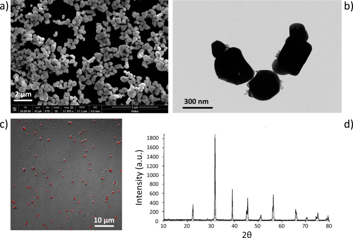
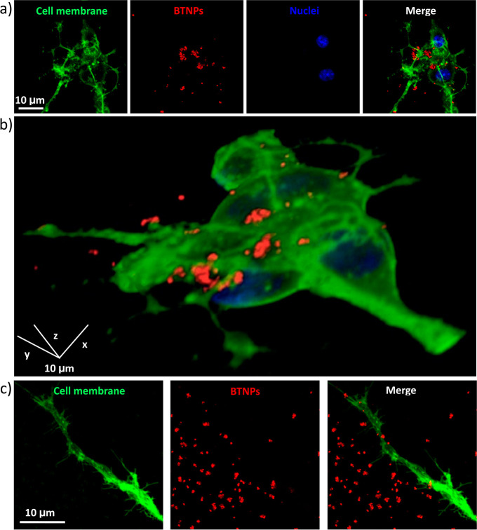
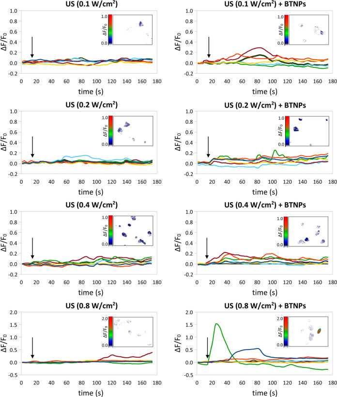
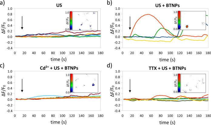
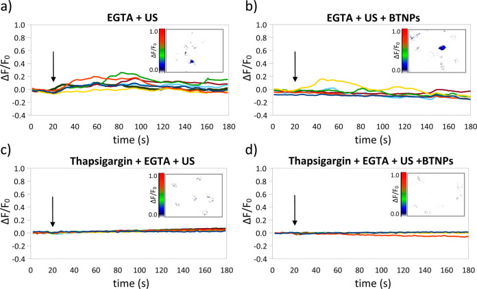
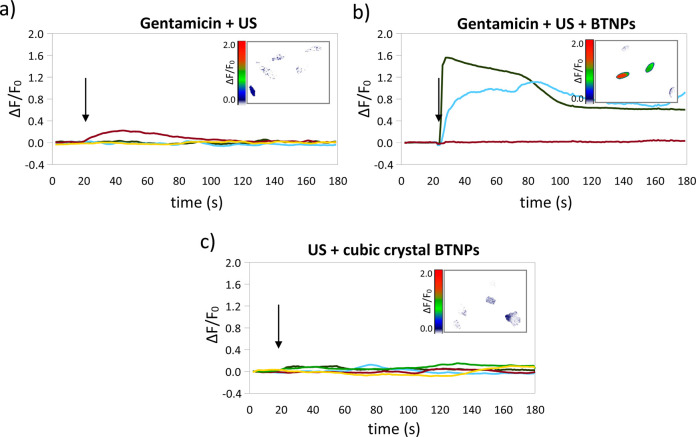
Tetragonal barium titanate nanoparticles (BTNPs) have been exploited as nanotransducers owing to their piezoelectric properties, in order to provide indirect electrical stimulation to SH-SY5Y neuron-like cells. Following application of ultrasounds to cells treated with BTNPs, fluorescence imaging of ion dynamics revealed that the synergic stimulation is able to elicit a significant cellular response in terms of calcium and sodium fluxes; moreover, tests with appropriate blockers demonstrated that voltage-gated membrane channels are activated. The hypothesis of piezoelectric stimulation of neuron-like cells was supported by lack of cellular response in the presence of cubic nonpiezoelectric BTNPs, and further corroborated by a simple electroelastic model of a BTNP subjected to ultrasounds, according to which the generated voltage is compatible with the values required for the activation of voltage-sensitive channels.

Keywords: SH-SY5Y cells; barium titanate nanoparticles; calcium imaging; piezoelectricity; ultrasounds.


Conflict of interest statement

The authors declare no competing financial interest.

Figures

Figure 1
Figure 1
Barium titanate nanoparticles (BTNPs) characterized by a tetragonal crystalline structure. SEM (a), TEM (b) and confocal fluorescence (c) imaging of BTNPs. In (c), confocal fluorescence image (red) is merged with the transmitted light image (gray) of the same field of view. The crystallographic structure revealed thanks to the XRD analysis shows two close peaks at 2θ = 44.85° and 2θ = 45.38°, specific of the tetragonal configuration of the crystal (d).
Figure 2
Figure 2
Confocal fluorescence microscopy of BTNPs associating to the neuronal plasma membranes. (a) Characteristic confocal z-stack of BTNP-treated SH-SY5Y-derived neurons (neuronal plasma membranes in green, BTNPs in red and nuclei in blue). (b) 3D rendering of the confocal z-stacks of the same field as in (a). BTNPs were also detected associating to the neurite membranes (c).
Figure 3
Figure 3
Calcium imaging of SH-SY5Y-derived neurons in response to the US stimulation performed at different intensities (0.1, 0.2, 0.4 and 0.8 W/cm2), with or without BNTPs: time courses of the ΔF/F0 traces. Arrows indicate the moment when the 5-s US pulse was initiated; in the inlet of each graph a representative calcium imaging time-lapse frame is reported (at t = 30).
Figure 4
Figure 4
US + BTNP stimulation (0.8 W/cm2) evokes Cd2+ and TTX-sensitive calcium transients. Representative ΔF/F0 traces relative to calcium imaging time-lapses of SH-SY5Y-derived neurons stimulated by US (a), US + BTNPs (b), US + BTNPs in the presence of Cd2+ (c) or TTX (d). Arrows indicate the moment when the 5-s US pulse was initiated; in the inlet of each graph a representative calcium imaging time-lapse frame is reported (at t = 50).
Figure 5
Figure 5
US + BTNP stimulation (0.8 W/cm2) induces TTX-sensitive sodium transients. Representative ΔF/F0 traces relative to sodium imaging time-lapses of SH-SY5Y-derived neurons stimulated by US (a), US + BTNPs (b), US + BTNPs in the presence of Cd2+ (c) or TTX (d). Arrows indicate the moment when the 5-s US pulse was initiated; in the inlet of each graph a representative sodium imaging time-lapse frame is reported (at t = 27).
Figure 6
Figure 6
Calcium sources involved during US and US + BTNP stimulations (0.8 W/cm2). Representative ΔF/F0 traces relative to calcium imaging time-lapses of SH-SY5Y-derived neurons in calcium-free conditions stimulated by US (a) and US + BTNPs (b) show in both case low-amplitude calcium transients. Observed transients were completely hindered by depleting the Ca2+ flux from the endoplasmic reticulum with thapsigargin before both the US (c) and US + BTNP (d) stimulations. Arrows indicate the moment when the 5-s US pulse was initiated; in the inlet of each graph a representative calcium imaging time-lapse frame is reported (at t = 50).
Figure 7
Figure 7
Calcium transients induced by US + BTNP stimulation (0.8 W/cm2) are mediated by the piezoelectricity of the nanoparticles. Representative ΔF/F0 traces relative to calcium imaging time-lapses of SH-SY5Y-derived neurons stimulated by US (a) and US + BTNPs (b) in the presence of gentamicin, a blocker of mechano-sensitive cation channels which does not affect the voltage-gated Ca2+ currents. In (c) the Ca2+ time course of neurons stimulated by US and nonpiezoelectric BTNPs, characterized by a cubic crystalline configuration. Arrows indicate the moment when the 5-s US pulse was initiated; in the inlet of each graph a representative calcium imaging time-lapse frame is reported (at t = 50).

References

    1. Chang J.-Y. Brain Stimulation for Neurological and Psychiatric Disorders, Current Status and Future Direction. J. Pharmacol. Exp. Ther. 2004, 309, 1–7. 10.1124/jpet.103.049718. - DOI - PubMed
    1. Deisseroth K. Optogenetics. Nat. Methods 2011, 8, 26–29. 10.1038/nmeth.f.324. - DOI - PMC - PubMed
    1. Gradinaru V.; Mogri M.; Thompson K. R.; Henderson J. M.; Deisseroth K. Optical Deconstruction of Parkinsonian Neural Circuitry. Science 2009, 324, 354–359. 10.1126/science.1167093. - DOI - PMC - PubMed
    1. Tye K. M.; Prakash R.; Kim S.-Y.; Fenno L. E.; Grosenick L.; Zarabi H.; Thompson K. R.; Gradinaru V.; Ramakrishnan C.; Deisseroth K. Amygdala Circuitry Mediating Reversible and Bidirectional Control of Anxiety. Nature 2011, 471, 358–362. 10.1038/nature09820. - DOI - PMC - PubMed
    1. Witten I. B.; Lin S.-C.; Brodsky M.; Prakash R.; Diester I.; Anikeeva P.; Gradinaru V.; Ramakrishnan C.; Deisseroth K. Cholinergic Interneurons Control Local Circuit Activity and Cocaine Conditioning. Science 2010, 330, 1677–1681. 10.1126/science.1193771. - DOI - PMC - PubMed

Publication types