

How Egg Case Proteins Can Protect Cuttlefish Offspring?
- PMID: 26168161
- PMCID: PMC4500399
- DOI: 10.1371/journal.pone.0132836
How Egg Case Proteins Can Protect Cuttlefish Offspring?
Abstract
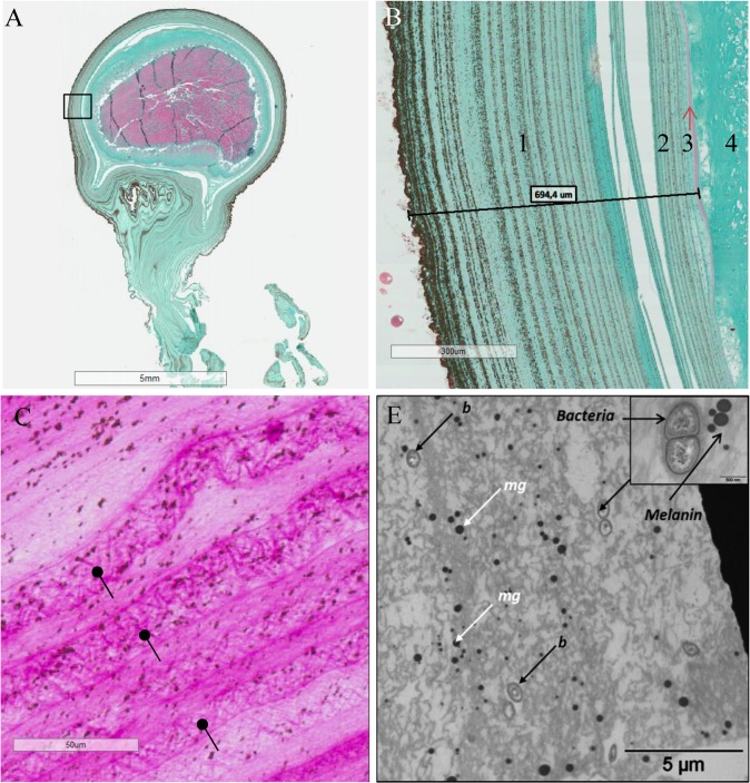
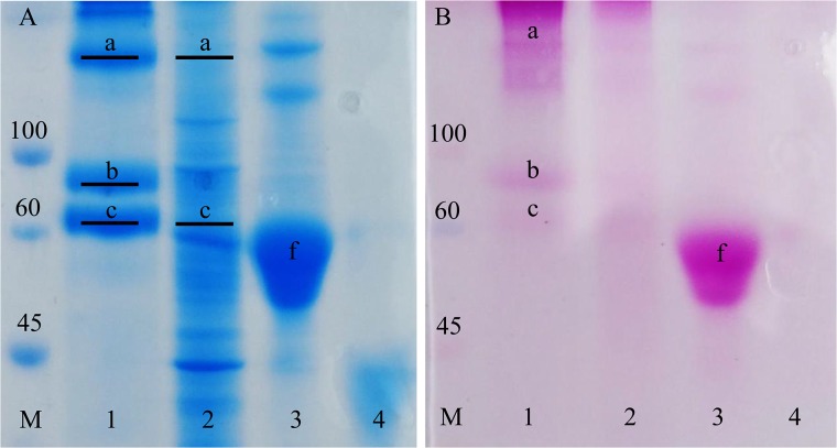
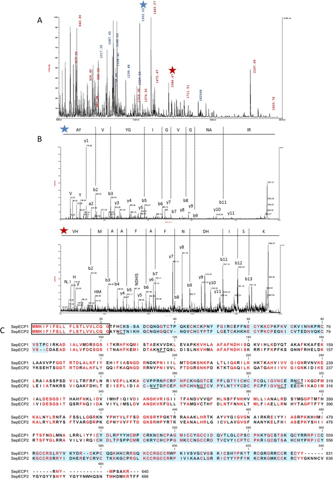
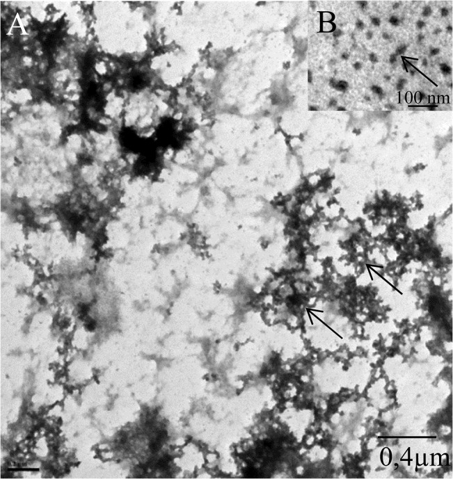
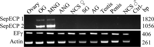
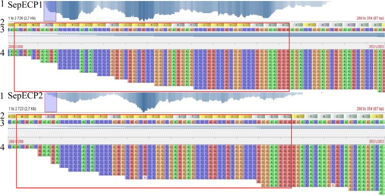
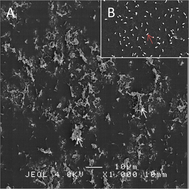
Sepia officinalis egg protection is ensured by a complex capsule produced by the female accessory genital glands and the ink bag. Our study is focused on the proteins constituting the main egg case. De novo transcriptomes from female genital glands provided essential databases for protein identification. A proteomic approach in SDS-PAGE coupled with MS unveiled a new egg case protein family: SepECPs, for Sepia officinalis Egg Case Proteins. N-glycosylation was demonstrated by PAS staining SDS-PAGE gels. These glycoproteins are mainly produced in the main nidamental glands. SepECPs share high sequence homology, especially in the signal peptide and the three cysteine-rich domains. SepECPs have a high number of cysteines, with conserved motifs involved in 3D-structure. SDS-PAGE showed that SepECPs could form dimers; this result was confirmed by TEM observations, which also revealed a protein network. This network is similar to the capsule network, and it associates these structural proteins with polysaccharides, melanin and bacteria to form a tight mesh. Its hardness and elasticity provide physical protection to the embryo. In addition, SepECPs also have bacteriostatic antimicrobial activity on GRAM- bacteria. By observing the SepECP / Vibrio aestuarianus complex in SEM, we demonstrated the ability of these proteins to agglomerate bacteria and thus inhibit their growth. These original proteins identified from the outer egg case ensure the survival of the species by providing physical and chemical protection to the embryos released in the environment without any maternal protection.
Conflict of interest statement
Figures
References
-
- Jonchère V, Réhault-Godbert S, Hennequet-Antier C, Cabau C, Sibut V, Cogburn LA, et al. Gene expression profiling to identify eggshell proteins involved in physical defense of the chicken egg. BMC Genomics [Internet]. 2010. January;11:57 Available: http://www.pubmedcentral.nih.gov/articlerender.fcgi?artid=2827412&tool=p.... Accessed 2014 Oct 31. 10.1186/1471-2164-11-57 - DOI - PMC - PubMed
-
- Guillette LJ, Fox SL, Palmer BD. Oviductal morphology and egg shelling in the oviparous lizardsCrotaphytus collaris andEumeces obsoletus. J Morphol [Internet]. 1989. August;201(2):145–59. Available: http://doi.wiley.com/10.1002/jmor.1052010205. Accessed 2014 Dec 1. - DOI - PubMed
-
- Marie P, Labas V, Brionne A, Harichaux G, Hennequet-Antier C, Nys Y, et al. Quantitative proteomics and bioinformatic analysis provide new insight into protein function during avian eggshell biomineralization. J Proteomics [Internet]. Elsevier B.V.; 2015. January 15;113:178–93. Available: http://www.ncbi.nlm.nih.gov/pubmed/25284052. Accessed 2014 Dec 17. 10.1016/j.jprot.2014.09.024 - DOI - PubMed
-
- Zhao A-C, Zhao T, Nakagaki K, Zhang Y, Sima Y, Miao Y, et al. Novel molecular and mechanical properties of egg case silk from wasp spider, Argiope bruennichi. Biochemistry [Internet]. 2006. March 14;45(10):3348–56. Available: http://www.ncbi.nlm.nih.gov/pubmed/16519529. Accessed 2015 Jan 8. - PubMed
-
- Hu X, Kohler K, Falick AM, Moore AMF, Jones PR, Sparkman OD, et al. Egg case protein-1. A new class of silk proteins with fibroin-like properties from the spider Latrodectus hesperus. J Biol Chem [Internet]. 2005. June 3;280(22):21220–30. Available: http://www.ncbi.nlm.nih.gov/pubmed/15797873. Accessed 2014 Dec 1. - PubMed
MeSH terms
LinkOut - more resources
Full Text Sources
Other Literature Sources
