. 2015 Aug 3;125(8):3063-76.
doi: 10.1172/JCI80055. Epub 2015 Jul 13.

T lymphocytes and fractalkine contribute to myocardial ischemia/reperfusion injury in patients

T lymphocytes and fractalkine contribute to myocardial ischemia/reperfusion injury in patients

Stephen E Boag et al. J Clin Invest. .

Abstract

Background: Lymphocytes contribute to ischemia/reperfusion (I/R) injury in several organ systems, but their relevance in ST elevation myocardial infarction (STEMI) is unknown. Our goal was to characterize lymphocyte dynamics in individuals after primary percutaneous coronary intervention (PPCI), assess the prognostic relevance of these cells, and explore mechanisms of lymphocyte-associated injury.

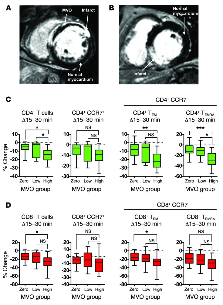
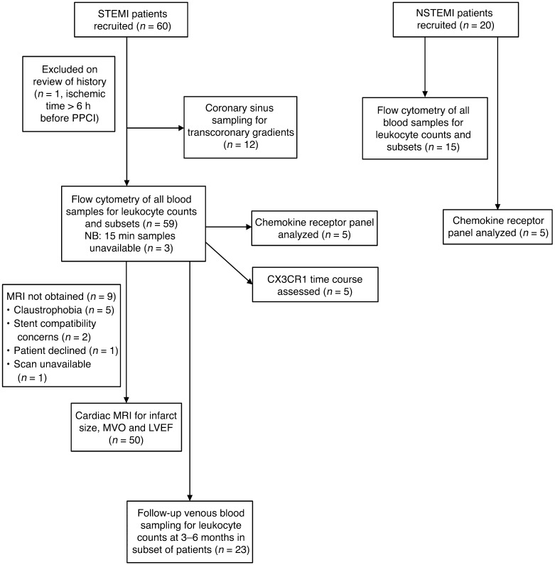
Methods: Lymphocyte counts were retrospectively analyzed in 1,377 STEMI patients, and the prognostic relevance of post-PPCI lymphopenia was assessed by Cox proportional hazards regression. Blood from 59 prospectively recruited STEMI patients undergoing PPCI was sampled, and leukocyte subpopulations were quantified. Microvascular obstruction (MVO), a component of I/R injury, was assessed using MRI.

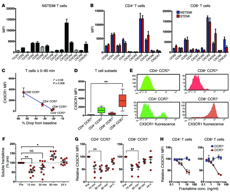
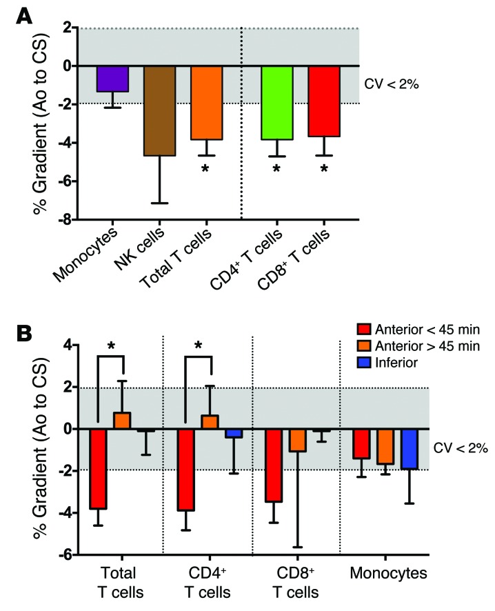
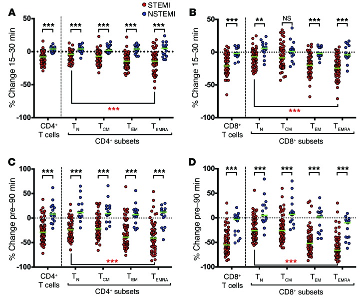
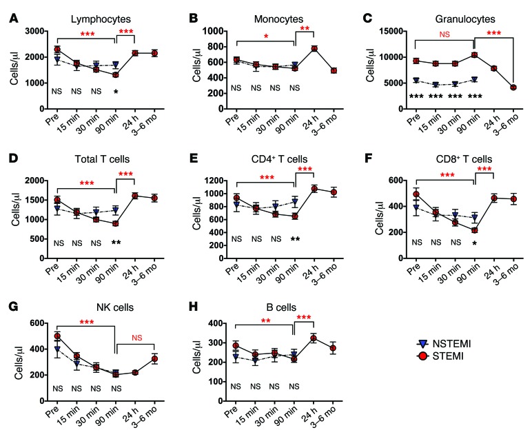
Results: In the retrospective cohort, lymphopenia was associated with a lower rate of survival at 3 years (82.8% vs. 96.3%, lowest vs. highest tertile; hazard ratio 2.42). In the prospective cohort, lymphocyte counts fell 90 minutes after reperfusion, primarily due to loss of T cells. CD8+ T cells decreased more than CD4+ T cells, and effector subsets exhibited the largest decline. The early decrease in effector T cell levels was greater in individuals that developed substantial MVO. The drop in T cell subsets correlated with expression of the fractalkine receptor CX3CR1 (r2 = 0.99, P = 0.006). Serum fractalkine concentration peaked at 90 minutes after reperfusion, coinciding with the T cell count nadir.

Conclusions: Lymphopenia following PPCI is associated with poor prognosis. Our data suggest that fractalkine contributes to lymphocyte shifts, which may influence development of MVO through the action of effector T cells.

Trial registration: Not applicable.

Funding: British Heart Foundation (FS/12/31/29533) and National Institute of Health Research (NIHR) Newcastle Biomedical Research Centre.


Figures

Figure 8
Figure 8. Example of gating strategy for eight-color flow cytometry assay.
Lymphocytes and monocytes were gated on their characteristic scatter patterns. Lymphocytes were classified based on CD3 expression to identify T cells, then divided into CD4+ and CD8+ T cells, prior to separation into their four main subsets of naive (TN), central memory (TCM), effector memory (TEM), and TEMRA cells. Each subset was also analyzed for expression of the differentiation marker CD27. CD3 lymphocytes were characterized as NK cells or B cells based on expression of CD16 and CD56. Monocytes were subdivided by expression of CD16.
Figure 7
Figure 7. Patterns in expression of the chemokine receptor CX3CR1 and its ligand, fractalkine, suggest a role in T cell dynamics after reperfusion.
(A) Chemokine receptor expression measured by MFI in T cells in NSTEMI patients (n = 5). Receptors eliminated at this stage indicated by asterisks and gray bars. (B) Chemokine receptor expression in CD4+ and CD8+ T cells in NSTEMI (n = 5) and STEMI (n = 5). (C) Pre-reperfusion expression of CX3CR1 in T cell subsets (n = 5 subset of STEMI group) correlates strongly with the mean drop in cells in STEMI patients (n = 59), according to linear regression and Pearson correlation coefficient. (D) Pre-reperfusion T cell surface expression of CX3CR1 in STEMI patients differs between subsets (Friedman test with Dunn’s multiple-comparisons test) (n = 5); box plots show median (central line), 25th and 75th percentiles (box limits), and range (error bars). (E) T cell subset CX3CR1 expression histograms (1 representative example of n = 5). (F) Serum levels of sFKN in STEMI patients, relative to pre-reperfusion level (one-way ANOVA, with Holm-Šídák multiple comparisons test) (n = 9). (G) Surface expression of CX3CR1 in CD4+CCR7 and CD8+CCR7 T cells (MFI relative to pre-reperfusion value). The results indicate an apparent drop in expression, which, given the findings in H, could also be due to ligand binding (Friedman test with Dunn’s multiple-comparisons test) (n = 5). (H) Preincubation of blood with recombinant fractalkine prior to CX3CR1 expression analysis indicates competition and reduced fluorescence on CCR7 (CX3CR1-expressing) T cells (MFI relative to baseline value) (n = 3). *P < 0.05, **P < 0.01. conc., concentration.
Figure 6
Figure 6. MVO is associated with greater drop in effector T cells.
STEMI patients underwent cardiac MRI to detect and quantify MVO. (A) Example of anterior MI with MVO. (B) Inferior infarct without MVO. STEMI patients were categorized into tertiles based on extent of MVO (zero: n = 17, low: n = 13, high: n = 17). (C) Change in cell counts between 15 and 30 minutes after reperfusion for total CD4+ T cells, CD4+CCR7+ (TN and TCM combined) cells, and CD4+CCR7 effector (TEM and TEMRA) subsets. (D) As above for total CD8+ T cells and subsets. Box plots display median (central line), 25th and 75th percentiles (limits of box), and 5th and 95th percentiles (error bars). Statistics refer to differences between MVO groups as indicated (Kruskal-Wallis test with Dunn’s multiple-comparisons test) (n = 47); *P < 0.05, **P < 0.01, ***P < 0.001.
Figure 5
Figure 5. Transcoronary gradients in cell counts, indicating loss of some cells across myocardial circulation.
Blood was taken simultaneously from the aortic root and CS at the end of PPCI in a subset of STEMI patients (n = 12). Of these, n = 9 were anterior STEMI (samples taken at <45 minutes after reperfusion in n = 6; >45 minutes after reperfusion in n = 3) and n = 3 were inferior STEMI (all sampled at <45 minutes). (A) Percentage change in cell counts of leukocyte subsets between aorta (Ao) and CS for anterior MI with sampling at <45 minutes (n = 6). Negative values indicate a drop across myocardial circulation. Statistics refer to Wilcoxon signed rank test of aorta versus CS counts for the indicated populations. (B) Impact of sample timing and infarct location on transcoronary gradients. Statistics refer to difference between anterior infarcts with sampling before (n = 6) or after (n = 3) 45 minutes (Mann-Whitney U test). Note: Mean troponin T for anterior infarcts with sampling <45 minutes: 4,814 ± 1,811 ng/l, anterior infarcts with sampling >45 minutes: 9,705 ± 2,409 ng/l, inferior infarcts: 5,462 ± 2,890 ng/l, excluding larger infarcts in the anterior sampling <45 minutes cases as a possible cause for these findings. *P < 0.05.
Figure 4
Figure 4. Percentage change in counts of circulating T cell subsets.
(A and B) Changes in T cell subset counts in STEMI (red) and NSTEMI (blue), shown as scatter plots of percentage change between 15 and 30 minutes, in (A) CD4+ T cells and subsets and (B) CD8+ T cells and subsets. (C and D) Changes in T cell subset counts between pre-reperfusion and 90 minutes in (C) CD4+ T cells and subsets and (D) CD8+ T cells and subsets. Median change is indicated by green lines. Upper statistics (black) refer to differences between groups (STEMI and NSTEMI) for each subset (Mann-Whitney U test); lower statistics (red) indicate differences between subsets in the STEMI group (Friedman test) (STEMI n = 59, NSTEMI n = 15). **P < 0.01, ***P < 0.001.
Figure 3
Figure 3. Time courses in circulating leukocyte subset counts.
(AC) Major leukocyte subsets of (A) lymphocytes, (B) monocytes, and (C) granulocytes, showing change in cell counts over time. Time points were measured from reperfusion in the STEMI group and from first culprit vessel instrumentation (or initial “Pre” blood sampling if no intervention occurred in the NSTEMI group. (DF) T lymphocyte cell counts, including (D) total T cells, (E) CD4+ T cells, and (F) CD8+ T cells. (G and H) CD3 (non-T) lymphocyte subset counts: (G) NK cells and (H) B cells. Upper statistics (red) refer to differences in counts between the indicated time points in the STEMI group (Friedman test, with Dunn’s multiple-comparisons test); while lower statistics (black) refer to difference between STEMI and NSTEMI at corresponding time points (Mann-Whitney U test) (STEMI n = 59, NSTEMI n = 15). *P < 0.05, **P < 0.01, ***P < 0.001.
Figure 2
Figure 2. Flow chart for prospective cohort.
Study groups recruited and investigations carried out in each.
Figure 1
Figure 1. Lymphopenia after PPCI predicts higher mortality after PPCI in STEMI.
(A) Kaplan-Meier survival curves of 1,377 consecutive patients discharged alive following PPCI (follow up time 1,200 days). Patients were divided into three tertiles according to the minimum peripheral blood lymphocyte counts obtained during their admission. (B) Forest plot with outcome of stepwise Cox regression analysis for death over follow-up time, with covariates including age, sex, minimum lymphocyte tertile, previous angina, and serum creatinine (see Supplemental Methods for the full list of covariates) (n = 1,076). (C) Minimum lymphocyte count, divided by age group (<65: n = 756, 65–80: n = 472, >80: n = 149) and (D) Difference in minimum lymphocyte counts between survivors and non-survivors following PPCI, according to 3 age groups, compared using one-way ANOVA; ***P < 0.001.

References

    1. Yellon DM, Hausenloy DJ. Myocardial reperfusion injury. N Engl J Med. 2007;357(11):1121–1135. doi: 10.1056/NEJMra071667. - DOI - PubMed
    1. Hausenloy DJ, Ong SB, Yellon DM. The mitochondrial permeability transition pore as a target for preconditioning and postconditioning. Basic Res Cardiol. 2009;104(2):189–202. doi: 10.1007/s00395-009-0010-x. - DOI - PubMed
    1. Vinten-Johansen J. Involvement of neutrophils in the pathogenesis of lethal myocardial reperfusion injury. Cardiovasc Res. 2004;61(3):481–497. doi: 10.1016/j.cardiores.2003.10.011. - DOI - PubMed
    1. Ito H. No-reflow phenomenon in patients with acute myocardial infarction: its pathophysiology and clinical implications. Acta Med Okayama. 2009;63(4):161–168. - PubMed
    1. Huang Y, Rabb H, Womer KL. Ischemia-reperfusion and immediate T cell responses. Cell Immunol. 2007;248(1):4–11. doi: 10.1016/j.cellimm.2007.03.009. - DOI - PMC - PubMed

Publication types

MeSH terms