

Non-puerperal mastitis masking pre-existing breast malignancy: importance of follow-up imaging
- PMID: 26169080
- PMCID: PMC4825209
- DOI: 10.14366/usg.15024
Non-puerperal mastitis masking pre-existing breast malignancy: importance of follow-up imaging
Abstract
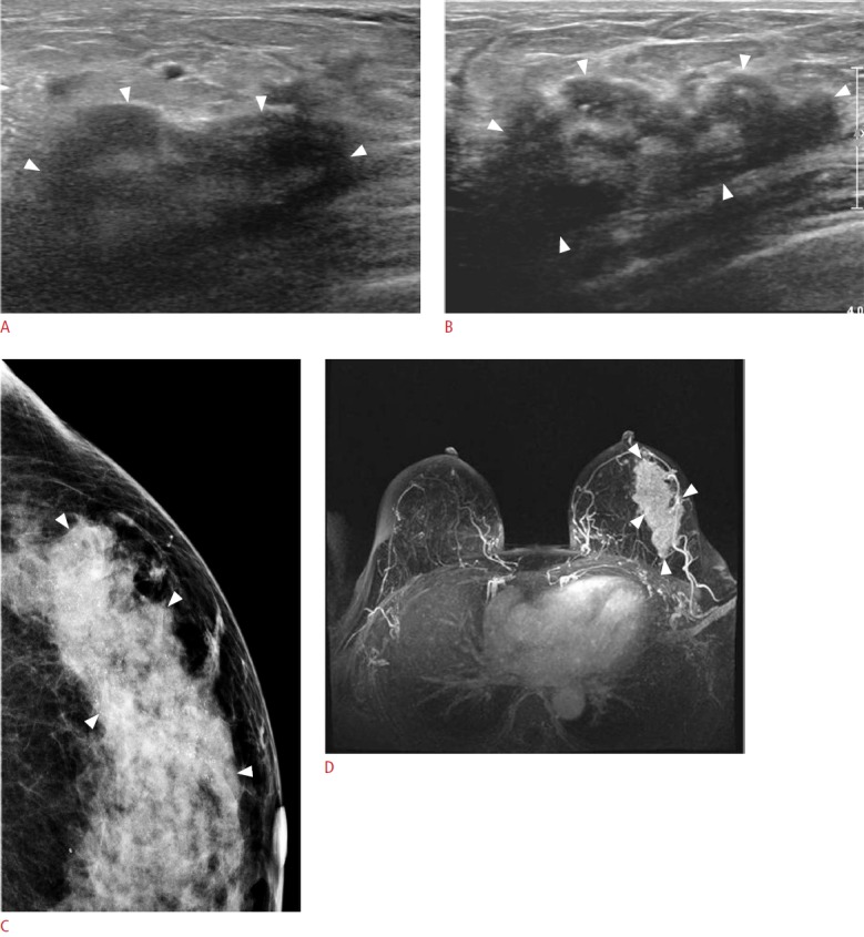
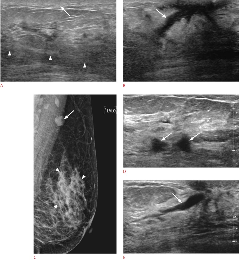
Mastitis is an inflammatory condition of the breast with common symptoms of pain, swelling, erythema, warmth, and fever. Diagnosis of mastitis is easily made on the basis of typical symptoms and ultrasonographic findings, such as diffusely increased echogenicity of the parenchyma and subcutaneous fat, or skin thickening. However, when it occurs in women middle-aged or older, associated malignancy should be considered. In our cases, we detected irregular hypoechoic malignant masses after the disappearance of inflammatory changes. Therefore, when non-puerperal women have inflammatory signs on their breast, follow-up imaging should be performed. In particular, in the case of persistent or growing palpability after the recovery of breast inflammation, percutaneous core biopsy and short-term follow-up with ultrasonography should be considered to exclude the associated malignancy.
Keywords: Breast; Breast neoplasms; Diagnosis; Mastitis; Ultrasonography.
Conflict of interest statement
No potential conflict of interest relevant to this article was reported.
Figures
References
-
- Boisserie-Lacroix M, Lafitte JJ, Sirben C, Latrabe V, Grelet P, Zeinoun R, et al. Inflammatory and infectious lesions of the breast: contribution of ultrasonography. J Chir (Paris) 1993;130:408–415. - PubMed
-
- Maier WP, Berger A, Derrick BM. Periareolar abscess in the nonlactating breast. Am J Surg. 1982;144:359–361. - PubMed
-
- Berna-Serna JD, Berna-Mestre JD. Follicular occlusion due to hyperkeratosis: a new hypothesis on the pathogenesis of mammillary fistula. Med Hypotheses. 2010;75:553–554. - PubMed
-
- Greydanus DE, Omar H, Pratt HD. The adolescent female athlete: current concepts and conundrums. Pediatr Clin North Am. 2010;57:697–718. - PubMed
-
- Page DL, Anderson TJ. Diagnostic histopathology of the breast. Edinburgh: Churchill Livingstone; 1987. pp. 64–65.
Publication types
LinkOut - more resources
Full Text Sources
Other Literature Sources
