

Cardiac fibroblasts: from development to heart failure
- PMID: 26169532
- PMCID: PMC4512919
- DOI: 10.1007/s00109-015-1314-y
Cardiac fibroblasts: from development to heart failure
Abstract
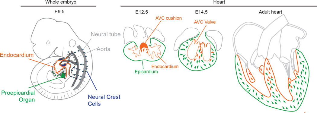
Cardiac fibroblasts are a major cell population of the heart and are characterized by their capacity to produce extracellular matrix (ECM). In hearts subjected to pressure overload, excessive fibroblast accumulation is responsible for fibrosis of the myocardium, a major clinical issue. Hence, understanding mechanisms generating fibroblasts in this context has become a key question in the cardiovascular field. Recent studies now point to the activation of resident fibroblasts as the underlying cause of fibrosis. However, de novo generation of fibroblasts from endothelium and circulating hematopoietic cells has also been proposed to significantly contribute to fibrosis. Here, we discuss the latest findings on fibroblast origins, with a particular emphasis on the pressure overload model, and the implication of these findings for the development of anti-fibrotic therapies that are currently lacking.
Figures

References
-
- Banerjee I, Yekkala K, Borg TK, Baudino TA. Dynamic interactions between myocytes, fibroblasts, and extracellular matrix. Annals of the New York Academy of Sciences. 2006;1080:76–84. - PubMed
-
- Porter KE, Turner NA. Cardiac fibroblasts: at the heart of myocardial remodeling. Pharmacology & therapeutics. 2009;123(2):255–278. - PubMed
-
- Mikawa T, Gourdie RG. Pericardial mesoderm generates a population of coronary smooth muscle cells migrating into the heart along with ingrowth of the epicardial organ. Developmental biology. 1996;174(2):221–232. - PubMed
-
- Gittenberger-de Groot AC, Vrancken Peeters MP, Mentink MM, Gourdie RG, Poelmann RE. Epicardium-derived cells contribute a novel population to the myocardial wall and the atrioventricular cushions. Circulation research. 1998;82(10):1043–1052. - PubMed
Publication types
MeSH terms
Grants and funding
LinkOut - more resources
Full Text Sources
Other Literature Sources
Medical
