. 2015 Aug;25(8):930-45.
doi: 10.1038/cr.2015.84. Epub 2015 Jul 14.

An HNF1α-regulated feedback circuit modulates hepatic fibrogenesis via the crosstalk between hepatocytes and hepatic stellate cells

Affiliations

An HNF1α-regulated feedback circuit modulates hepatic fibrogenesis via the crosstalk between hepatocytes and hepatic stellate cells

Hui Qian et al. Cell Res. 2015 Aug.

Abstract

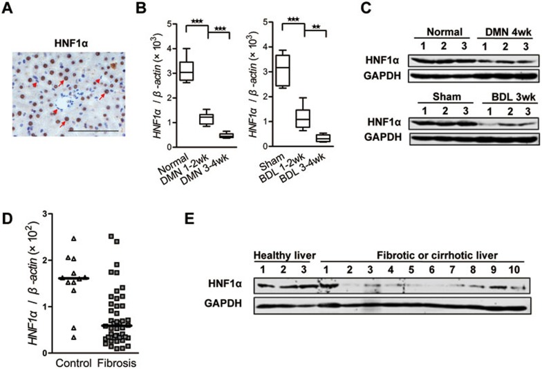
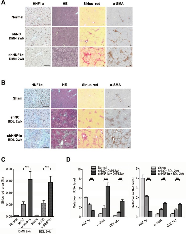
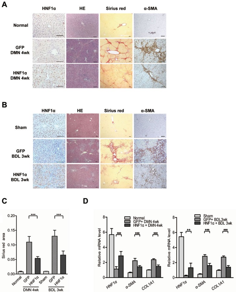
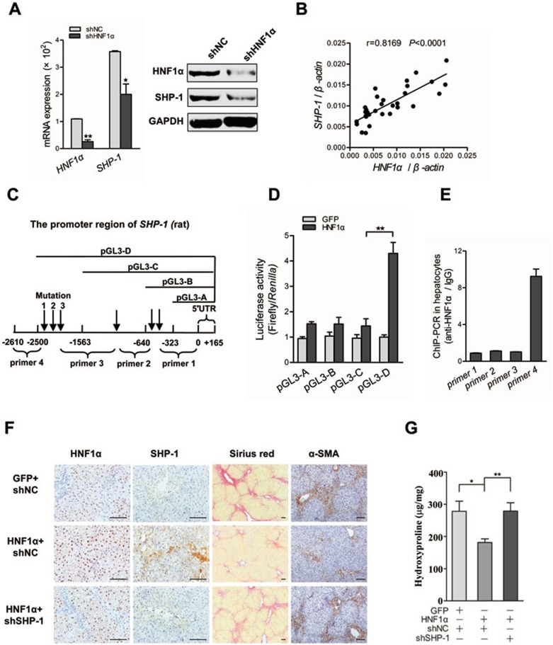
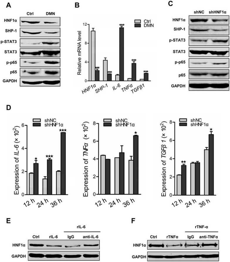
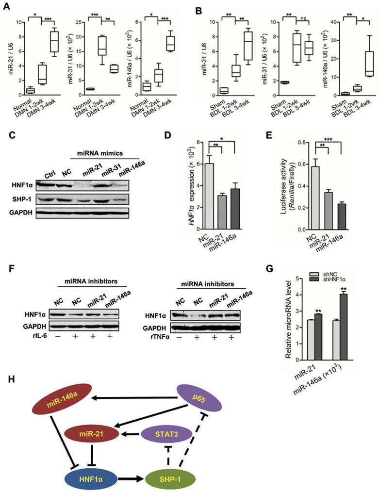
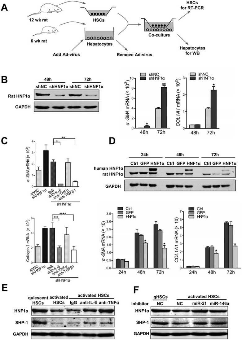
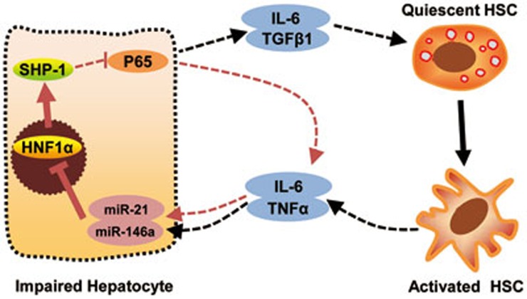
Hepatocytes are critical for the maintenance of liver homeostasis, but its involvement in hepatic fibrogenesis remains elusive. Hepatocyte nuclear factor 1α (HNF1α) is a liver-enriched transcription factor that plays a key role in hepatocyte function. Our previous study revealed a significant inhibitory effect of HNF1α on hepatocellular carcinoma. In this study, we report that the expression of HNF1α is significantly repressed in both human and rat fibrotic liver. Knockdown of HNF1α in the liver significantly aggravates hepatic fibrogenesis in either dimethylnitrosamine (DMN) or bile duct ligation (BDL) model in rats. In contrast, forced expression of HNF1α markedly alleviates hepatic fibrosis. HNF1α regulates the transcriptional expression of SH2 domain-containing phosphatase-1 (SHP-1) via directly binding to SHP-1 promoter in hepatocytes. Inhibition of SHP-1 expression abrogates the anti-fibrotic effect of HNF1α in DMN-treated rats. Moreover, HNF1α repression in primary hepatocytes leads to the activation of NF-κB and JAK/STAT pathways and initiates an inflammatory feedback circuit consisting of HNF1α, SHP-1, STAT3, p65, miR-21 and miR-146a, which sustains the deregulation of HNF1α in hepatocytes. More interestingly, a coordinated crosstalk between hepatocytes and hepatic stellate cells (HSCs) participates in this positive feedback circuit and facilitates the progression of hepatocellular damage. Our findings demonstrate that impaired hepatocytes play an active role in hepatic fibrogenesis. Early intervention of HNF1α-regulated inflammatory feedback loop in hepatocytes may have beneficial effects in the treatment of chronic liver diseases.


Figures

Figure 1
Figure 1
HNF1α is repressed in fibrotic liver. (A) Immunohistochemical staining of HNF1α in normal rat liver. HNF1α is detected exclusively in the nuclei of hepatocytes (arrow). No obvious staining is observed in non-parenchymal cells (arrow head). Scale bar, 100 μm. (B) mRNA level of HNF1α was assessed by real-time PCR in the livers treated with dimethylnitrosamine (DMN, left) or bile duct ligation (BDL, right) (n = 6 in each group). **P < 0.01; ***P < 0.001 by Mann-Whitney U test. (C) HNF1α protein level in the liver of 3 individual rats after DMN injection (top) or BDL operation (bottom) was detected. (D) A scatter dot plot showing HNF1α expression levels in 12 human control and 44 fibrotic samples as assessed by RT-PCR analysis. Data (median) are normalized to β-actin, and P value was computed by Mann-Whitney U test (P = 0.0008). (E) Western blot analysis of HNF1α in the livers from 3 healthy control individuals and 10 patients with either fibrosis or cirrhosis.
Figure 2
Figure 2
Repression of HNF1α aggravates hepatic fibrogenesis in both DMN and BDL models. (A, B) Adenovirus carrying shRNA against HNF1α (shHNF1α) or negative control (shNC) was injected into rats prior to DMN administration (A) and BDL treatment (B), and 2 weeks later the expression of HNF1α and α-SMA in the fibrotic livers was analyzed by immunohistochemistry. Hematoxylin and eosin (HE) and Sirius red staining were used to examine pathological alterations and collagen deposition. (C) Semi-quantitative analysis of Sirius red staining in the fibrotic livers from AdshHNF1α or AdshNC-treated rats (n = 10 rats in each group). (D) mRNA levels of HNF1α, α-SMA and COL1A1 in the livers were detected by real-time PCR Scale bar, 100 μm. **P < 0.01; ***P < 0.001.
Figure 3
Figure 3
HNF1α overexpression attenuates hepatic fibrosis. (A, B) A single dose of adenovirus carrying human HNF1α gene (HNF1α) or control virus (GFP) was injected into rats after DMN injection (A) or BDL operation (B). The fibrotic livers were analyzed at 4 weeks after DMN treatment or 3 weeks after BDL. The expression of HNF1α and α-SMA was assessed by immunohistochemistry. HE and Sirius red staining were used to examine pathological alterations and collagen deposition. (C) Semi-quantitative analysis of Sirius red staining in the fibrotic livers from AdHNF1α or AdGFP-treated rats (n = 6 rats in each group). (D) mRNA levels of HNF1α, α-SMA and COL1A1 in the livers were detected by real-time PCR. Scale bars, 100 μm. **P < 0.01; ***P < 0.001.
Figure 4
Figure 4
Anti-fibrotic effect of HNF1α depends on the transcriptional activation of SHP-1. (A) Transcript level of HNF1α and SHP-1 in primary rat hepatocytes treated with AdshHNF1α or AdshNC. (B) Correlation between the mRNA levels of HNF1α and SHP-1 in human liver tissues. Each data point represents an individual sample, and the correlation coefficient (r) is shown. (C) A schematic representation of the promoter region of SHP-1, the potential cis-acting elements for HNF1α (arrow), mutation sites and the fragment amplified in ChIP-PCR. (D) The nested deletion analysis shows the transactivation effect of HNF1α on rat SHP-1 promoter. (E) HNF1α occupancy at the SHP-1 loci detected by ChIP-PCR in freshly isolated hepatocytes. (F) Suppression of SHP-1 reverses the anti-fibrotic effect of HNF1α. AdshSHP-1 or AdshNC was simultaneously delivered with AdHNF1α into DMN-treated rats. Collagen deposition and the expression of HNF1α, SHP-1 and α-SMA were detected in the livers. (G) Hydroxyproline content was assayed in the fibrotic livers (n = 9 rats in each group). Scale bars, 100 μm. *P < 0.05; **P < 0.01; ***P < 0.001.
Figure 5
Figure 5
HNF1α suppression aggravates hepatocellular inflammation. (A) Western blot analysis of HNF1α, SHP-1 and phosphorylation of STAT3 and p65 in the lysates of hepatocytes isolated from rats with DMN treatment for 2 weeks. (B) mRNA levels of HNF1α, SHP-1, IL-6, TNFα and TGFβ1 in hepatocytes from rats with DMN treatment for 2 weeks vs the control rats. (C) Representative western blot of HNF1α, SHP-1, p65 and STAT3 in hepatocytes treated with AdshHNF1α or AdshNC. (D) Transcript levels of IL-6, TNFα and TGFβ1 in hepatocytes treated with AdshHNF1α or AdshNC for 12-36 h. (E, F) Protein level of HNF1α in the hepatocytes stimulated by recombinant IL-6 (rIL-6, 50 ng/ml, E) or recombinant TNFα (rTNFα, 20 ng/ml, F). Rabbit antibody against IL-6 (anti-IL-6) or against TNFα (anti-TNFα) was simultaneously added into medium to block the effect of IL-6 or TNFα, respectively. Normal rabbit IgG was used as control. *P < 0.05; **P < 0.01; ***P < 0.001.
Figure 6
Figure 6
An HNF1α-regulated inflammatory circuit mediates hepatocellular impairment. (A, B) Real-time PCR analysis of miR-21, miR-31 and miR-146a in liver tissues from rats with DMN injection (n = 6 in each group) (A) and BDL treatment (n = 6 in each group) (B). (C) Western blot analysis of HNF1α and SHP-1 in hepatocytes transfected with indicated miRNA mimics for 72 h. (D) mRNA level of HNF1α in hepatocytes transfected with miRNA mimics for 48 h. (E) The effect of miR-21 and miR-146a mimics on luciferase activity of HNF1α 3′ UTR in HEK293T cells. (F) Western blot analysis of HNF1α in hepatocytes transfected with miRNA inhibitors and treated with IL-6 (rIL-6, left) or TNFα (rTNFα, right) for 48 h. (G) Levels of miR-21 and miR-146a in hepatocytes treated with AdshHNF1α or AdshNC. (H) A schematic model of the proposed HNF1α feedback circuit in hepatocellular damage. *P < 0.05; **P < 0.01; ***P < 0.001.
Figure 7
Figure 7
Crosstalk between HSCs and hepatocytes in vitro. (A) A schematic representation of co-culture experiments with primary HSCs and hepatocytes isolated from rats. (B) Suppression of HNF1α in hepatocytes enhances the activation of HSCs. Endogenous HNF1α level in primary rat hepatocytes pretreated with AdshHNF1α or AdshNC was detected by western blot (left). mRNA levels of α-SMA and COL1A1 in HSCs were assessed by RT-PCR (right). (C) mRNA level of α-SMA and COL1A1 in HSCs co-cultured with AdshHNF1α- or AdshNC-treated hepatocytes. Antibody against IL-6, TNFα or TGFβ1 was added into the co-culture to block the corresponding cytokine. (D) Hepatocytes overexpressing HNF1α attenuates the activation of HSCs. Expression of exogenous human HNF1α and endogenous rat HNF1α in hepatocytes treated with AdHNF1α or AdGFP analyzed by western blot is shown in the top panel; mRNA levels of α-SMA and COL1A1 in HSCs are shown in the bottom panels. (E) Western blot analysis of HNF1α and SHP-1 in hepatocytes co-cultured with quiescent or activated HSCs for 48 h. Antibodies against TNFα, IL-6 and control IgG were used to block the cytokines in co-culture. (F) Expression of HNF1α and SHP-1 in hepatocytes transfected with miRNA inhibitors and then co-cultured with quiescent or activated HSCs for 48 h.
Figure 8
Figure 8
An HNF1α-mediated feedback circuit modulates the crosstalk between HSCs and hepatocytes. A schematic presentation of the proposed autocrine regulation and crosstalk between HSCs and hepatocytes. An intrinsic inflammatory feedback loop could aggravate the hepatocellular impairment. Inhibition of HNF1α in hepatocytes by miR-21 and miR-146a leads to an increase of IL-6 and TGFβ1 production, which causes the activation of HSCs. On the other hand, activated HSCs secrete IL-6 and TNFα, which further suppress the expression of HNF1α and SHP-1 in hepatocytes.

References

    1. Bataller R, Brenner DA. Liver fibrosis. J Clin Invest. 2005;115:209–218. - PMC - PubMed
    1. Schuppan D, Afdhal NH. Liver cirrhosis. Lancet. 2008;371:838–851. - PMC - PubMed
    1. Friedman SL. Mechanisms of hepatic fibrogenesis. Gastroenterology. 2008;134:1655–1669. - PMC - PubMed
    1. Hernandez-Gea V, Friedman SL. Pathogenesis of liver fibrosis. Annu Rev Pathol. 2011;6:425–456. - PubMed
    1. Friedman SL. Evolving challenges in hepatic fibrosis. Nat Rev Gastroenterol Hepatol. 2010;7:425–436. - PubMed

Publication types

LinkOut - more resources