. 2015 Aug 4;54(30):4665-71.
doi: 10.1021/acs.biochem.5b00608. Epub 2015 Jul 27.

Tissue Factor Residues That Modulate Magnesium-Dependent Rate Enhancements of the Tissue Factor/Factor VIIa Complex

Affiliations

Tissue Factor Residues That Modulate Magnesium-Dependent Rate Enhancements of the Tissue Factor/Factor VIIa Complex

Joshua M Gajsiewicz et al. Biochemistry. .

Abstract

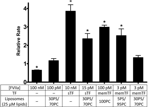
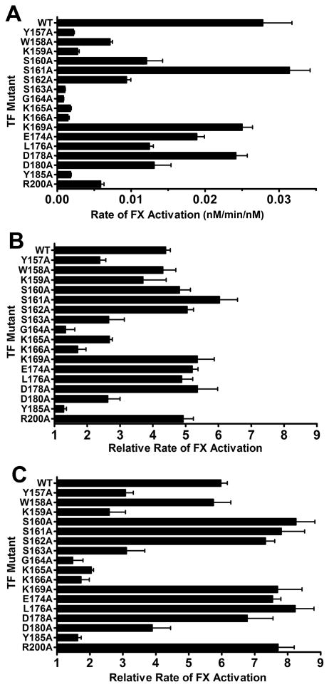
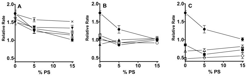
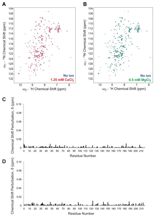
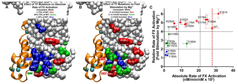
The blood coagulation cascade is initiated when the cell-surface complex of factor VIIa (FVIIa, a trypsin-like serine protease) and tissue factor (TF, an integral membrane protein) proteolytically activates factor X (FX). Both FVIIa and FX bind to membranes via their γ-carboxyglutamate-rich domains (GLA domains). GLA domains contain seven to nine bound Ca(2+) ions that are critical for their folding and function, and most biochemical studies of blood clotting have employed supraphysiologic Ca(2+) concentrations to ensure saturation of these domains with bound Ca(2+). Recently, it has become clear that, at plasma concentrations of metal ions, Mg(2+) actually occupies two or three of the divalent metal ion-binding sites in GLA domains, and that these bound Mg(2+) ions are required for full function of these clotting proteins. In this study, we investigated how Mg(2+) influences FVIIa enzymatic activity. We found that the presence of TF was required for Mg(2+) to enhance the rate of FX activation by FVIIa, and we used alanine-scanning mutagenesis to identify TF residues important for mediating this response to Mg(2+). Several TF mutations, including those at residues G164, K166, and Y185, blunted the ability of Mg(2+) to enhance the activity of the TF/FVIIa complex. Our results suggest that these TF residues interact with the GLA domain of FX in a Mg(2+)-dependent manner (although effects of Mg(2+) on the FVIIa GLA domain cannot be ruled out). Notably, these TF residues are located within or immediately adjacent to the putative substrate-binding exosite of TF.


Figures

Figure 1
Figure 1
Effect of Mg2+ on FX activation by FVIIa in the presence or absence of TF or membranes. FX activation by FVIIa was measured in the presence of the components listed, with a dash (—) indicating the component was not present. Data are mean initial rates of FX activation in the presence of 1.25 mM Ca2+ and 0.6 mM Mg2+ normalized to rates using 1.25 mM Ca2+ alone. Error bars are one standard error (n ≥ 3). Normalized rates that are statistically significantly different from 1.0 are indicated with an asterisk (one-sample t-tests; p < 0.05).
Figure 2
Figure 2
Effect of TF mutations on rates of FX activation by the memTF/FVIIa complex in solution (with 0.1% Triton X-100), measured using different divalent metal ion concentrations. (A) FX activation by the memTF/FVIIa complex using 1.85 mM Ca2+ alone, graphed as initial rates of FX activation divided by the memTF/FVIIa complex concentration. (B) Relative rates of FX activation by memTF/FVIIa in solution using 1.25 mM Ca2+ and 0.6 mM Mg2+, normalized to rates using 1.85 mM Ca2+ alone. (C) Relative rates of FX activation by the memTF/FVIIa complex in solution using 1.25 mM Ca2+ and 0.6 mM Mg2+, normalized to rates using 1.25 mM Ca2+ alone. Data are means ± the standard error (n ≥ 3).
Figure 3
Figure 3
Effect of 11 selected TF mutations on the ability of Mg2+ to enhance the rate of FX activation by TF/FVIIa/membrane complexes. WT or mutant memTF was incorporated into liposomes containing 0-15% PS, with the balance being PC. Initial rates of FX activation by the resulting memTF/FVIIa complexes were measured with 1.25 mM Ca2+ and 0.6 mM Mg2+ and normalized to the rates observed with 1.85 mM Ca2+ alone. The memTF mutants are grouped in the three panels as described in the text, with the same data for WT memTF (●) plotted in each panel for comparison. (A) Normalized rates of FX activation observed with memTF mutants S162A (⨯), W158A (▼), E174A (∇), and D178A (✯). (B) Normalized rates of FX activation observed with memTF mutants S163A (□), K165A (◆), K159A (○), and Y157A (▲). (C) Normalized rates of FX activation observed with memTF mutants Y185A (∆), K166A (■), and G164A (◊). Data are means ± the standard error (n ≥ 3).
Figure 4
Figure 4
Lack of a detectable effect of divalent metal ions on NMR spectra of sTF. (A) 15N-1H 2D HSQC correlation spectrum of 100 μM sTF in 50 mM sodium phosphate, 50 mM NaCl, 1 mM DSS, 10% D2O and no divalent metal ions (blue) overlaid with the sTF spectrum in the same buffer with 1.25 mM Ca2+ (red). (B) 15N-1H 2D HSQC correlation spectrum of 100 μM sTF in the same buffer as in panel A, without divalent metal ions (blue) overlaid with the spectrum in the presence of 0.5 mM Mg2+ (green). (C) Chemical shift perturbations upon titration with 1.25 mM Ca2+ (from panel A) calculated as δ = ([0.1δN]2 + δH2)1/2), with an average chemical shift perturbation of 0.0014 ppm. (D) Chemical shift perturbations upon titration with 0.5 mM Mg2+ (from panel B), with an average chemical shift perturbation of 0.0015 ppm. Of the 208 expected non-proline resonances in sTF, 196 resonances were assigned.
Figure 5
Figure 5
Localization and properties of TF residues investigated. The structure is from Protein Data Bank entry 3TH2, in which sTF/FVIIa was crystallized in the presence of 5 mM Ca2+ and 2.5 mM Mg2+. FVIIa is depicted as orange ribbons, with bound Ca2+ ions colored teal and Mg2+ ions beige. (A) Localization of the TF residues tested in this study for their effect on the absolute rate of FX activation by memTF/FVIIa in solution. TF residues are color-coded according to their rate of FX activation in the presence of 1.85 mM Ca2+ alone (from Figure 2A). Unmutated TF residues are colored white. TF residues are colored red, which, when mutated, retained essentially WT activity (i.e., ≥ 75% of the WT rate of FX activation). Residues with a moderate effect on the FX activation rate are colored green (30-75% of the WT rate); residues with severe defects are colored blue (≤ 30% of the WT rate). Residues W158 and S160, obstructed in this view, are colored blue and green, respectively. (B) Localization of TF residues tested in this study as being important for the ability of Mg2+ to enhance the rate of FX activation by memTF/FVIIa in solution. TF residues are color-coded according to their rate of FX activation in the presence of Ca2+ and Mg2+ normalized to the rate at 1.85 mM Ca2+ alone (from Figure 2B). Unmutated TF residues are colored white. TF residues are colored red, which, when mutated, retained essentially WT responses to Mg2+ (i.e., 4-6-fold enhancement of the FX activation rate). Residues with blunted responses to Mg2+ are colored green (2-4-fold enhancement by Mg2+) or blue (1-2-fold enhancement by Mg2+). Residues W158 and S160, obstructed in this view are colored red. (C) Relative rate of FX activation (rate with 1.25 mM Ca2+ and 0.6 mM Mg2+, divided by the rate with 1.85 mM Ca2+) vs the absolute rate of FX activation, in both cases by memTF/FVIIa in solution (i.e., data replotted from panels A and B of Figure 2, respectively). Residues are color-coded as in panel B. Vertical black dotted lines correspond to the color-coding cutoff values from panel A; horizontal black dotted lines correspond to the color-coding cutoff values from panel B. A vertical dashed red line marks the location of WT TF.

References

    1. Vadivel K, Agah S, Messer AS, Cascio D, Bajaj MS, Krishnaswamy S, Esmon CT, Padmanabhan K, Bajaj SP. Structural and functional studies of γ-carboxyglutamic acid domains of factor VIIa and activated protein C: Role of magnesium at physiological calcium. J Mol Biol. 2013;425:1961–1981. - PMC - PubMed
    1. Bloom JW, Mann KG. Metal ion induced conformational transitions of prothrombin and prothrombin fragment 1. Biochemistry. 1978;17:4430–4438. - PubMed
    1. Zwaal RFA, Comfurius P, Bevers EM. Lipid-protein interactions in blood coagulation. Biochim Biophys Acta. 1998;1376:433–453. - PubMed
    1. Sunnerhagen M, Forsén S, Hoffrén AM, Drakenberg T, Teleman O, Stenflo J. Structure of the Ca2+-free GLA domain sheds light on membrane binding of blood coagulation proteins. Nat Struct Biol. 1995;2:504–509. - PubMed
    1. Falls LA, Furie BC, Jacobs M, Furie B, Rigby AC. The ω-loop region of the human prothrombin γ-carboxyglutamic acid domain penetrates anionic phospholipid membranes. J Biol Chem. 2001;276:23895–23902. - PubMed

Publication types

MeSH terms

LinkOut - more resources