. 2015 Jul 27;212(8):1267-81.
doi: 10.1084/jem.20142358. Epub 2015 Jul 13.

Blocking neutrophil integrin activation prevents ischemia-reperfusion injury

Affiliations

Blocking neutrophil integrin activation prevents ischemia-reperfusion injury

Tadayuki Yago et al. J Exp Med. .

Abstract

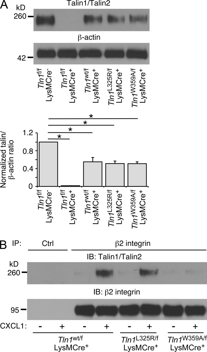
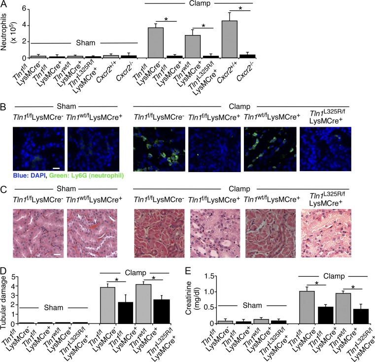
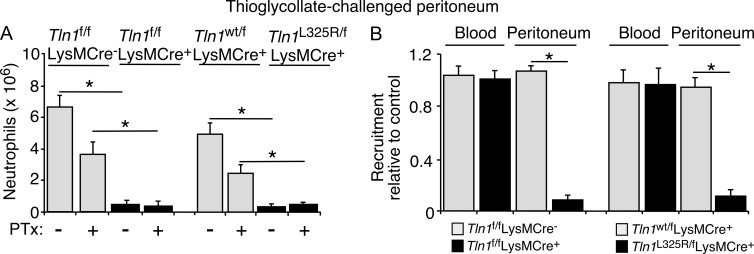
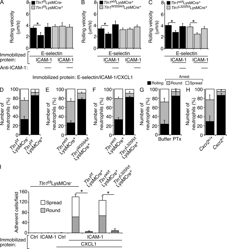
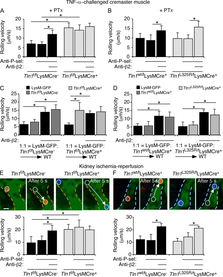
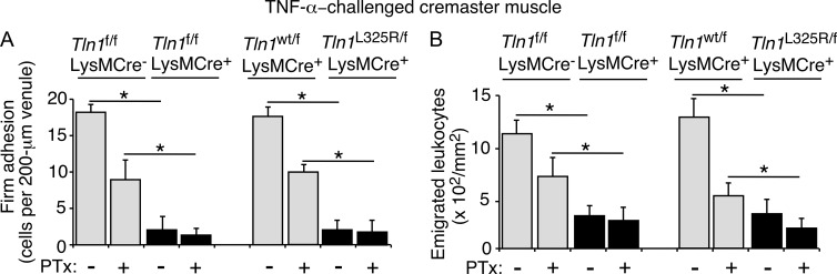
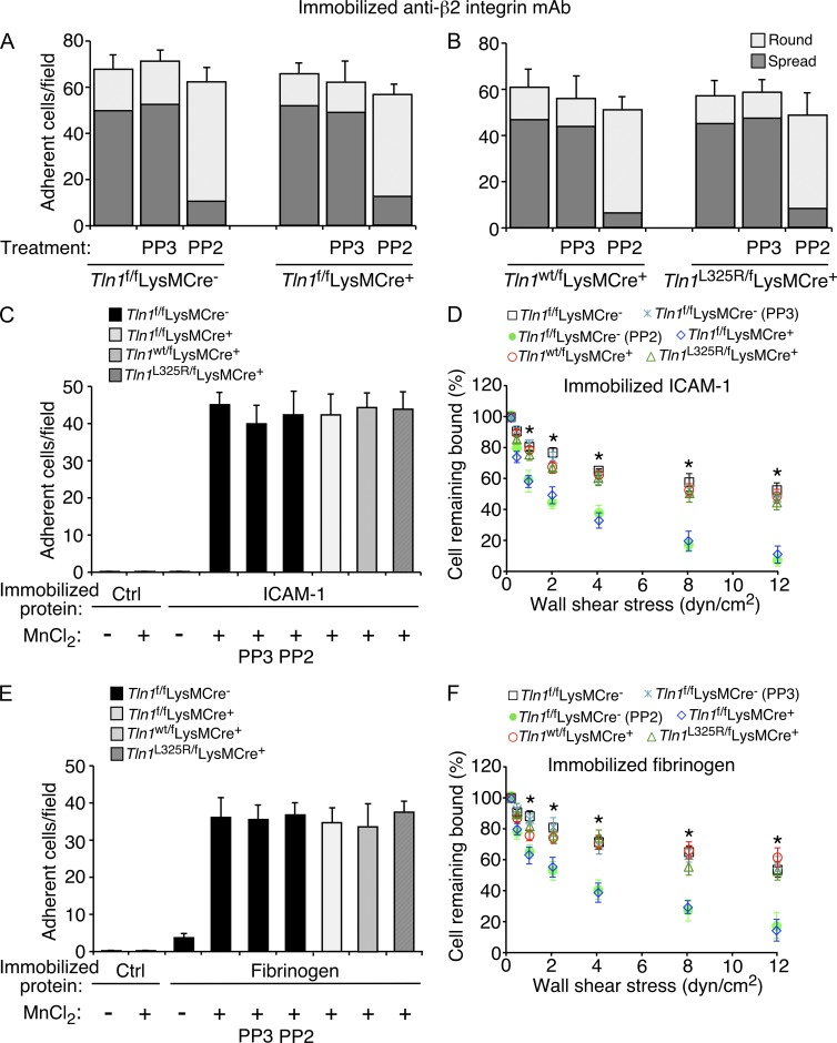
Neutrophil recruitment, mediated by β2 integrins, combats pyogenic infections but also plays a key role in ischemia-reperfusion injury and other inflammatory disorders. Talin induces allosteric rearrangements in integrins that increase affinity for ligands (activation). Talin also links integrins to actin and other proteins that enable formation of adhesions. Structural studies have identified a talin1 mutant (L325R) that perturbs activation without impairing talin's capacity to link integrins to actin and other proteins. Here, we found that mice engineered to express only talin1(L325R) in myeloid cells were protected from renal ischemia-reperfusion injury. Dissection of neutrophil function in vitro and in vivo revealed that talin1(L325R) neutrophils had markedly impaired chemokine-induced, β2 integrin-mediated arrest, spreading, and migration. Surprisingly, talin1(L325R) neutrophils exhibited normal selectin-induced, β2 integrin-mediated slow rolling, in sharp contrast to the defective slow rolling of neutrophils lacking talin1 or expressing a talin1 mutant (W359A) that blocks talin interaction with integrins. These studies reveal the importance of talin-mediated activation of integrins for renal ischemia-reperfusion injury. They further show that neutrophil arrest requires talin recruitment to and activation of integrins. However, although neutrophil slow rolling requires talin recruitment to integrins, talin-mediated integrin activation is dispensable.


Figures

Figure 1.
Figure 1.
Murine neutrophils express equivalent levels of talin1, talin1(L325R), or talin1(W359A). (A, top) Representative Western blot of neutrophil lysates from the indicated genotype, probed with mAb 8d4, which recognizes both talin1 and talin2, or probed with rabbit anti–β-actin IgG. (bottom) Quantification of the talin/β-actin ratio by densitometry. (B) Neutrophils of the indicated genotype were incubated with or without CXCL1, lysed, and immunoprecipitated (IP) with control (Ctrl) or anti–β2 integrin mAb. Immunoprecipitates were analyzed by Western blotting (immunoblot, IB) with anti-talin and anti–β2 integrin antibodies. The data in A represent the mean ± SEM from three experiments. The data in B are representative of three experiments. *, P < 0.01, as determined by unpaired Student’s t test.
Figure 2.
Figure 2.
Mice expressing talin1(L325R) in neutrophils are protected from kidney ischemia–reperfusion injury. Mice of the indicated genotype were subjected to sham surgery or surgery that included a 30-min clamp of both renal pedicles to induce ischemia, followed by release of the clamp to permit reperfusion for 24 h. (A) Neutrophils infiltrating the kidneys were quantified by flow cytometry. (B) Immunofluorescence of representative kidney sections. Neutrophils were stained with anti-Ly6G mAb. Nuclei were counterstained with DAPI. Bar, 10 µm. (C) Representative kidney outer medulla sections stained with hematoxylin and eosin. Bar, 50 µm. (D) Tubular necrosis was quantified by epithelial karyolysis, necrotic debris, and cast formation as described in Materials and methods. (E) Creatinine levels in plasma. At least five mice were in each experimental group. The data in B and C are representative of five experiments. The data in A, D, and E represent the mean ± SEM from five experiments. *, P < 0.01, as determined by unpaired Student’s t test.
Figure 3.
Figure 3.
Neutrophils expressing talin1(L325R) exhibit impaired entry into a site of inflammation. (A) Untreated or PTx-pretreated mice of the indicated genotype were injected with thioglycollate intraperitoneally. After 4 h, peritoneal cells were collected, and the number of neutrophils was measured by flow cytometry. (B) Bone marrow leukocytes from control mice (Tln1f/fLysMCre or Tln1f/wtLysMCre+) or experimental mice (Tln1f/fLysMCre+ or Tln1f/L325RLysMCre+) were labeled with red (PKH26) or green (PKH67) dye. The labeled cells were resuspended at 108 cells/ml and mixed at a 1:1 ratio. Recipient mice were injected with thioglycollate intraperitoneally and, after 2 h, with 200 µl of the labeled cell mixture retroorbitally. After another 2 h, blood was collected. The mice were sacrificed, and peritoneal cells were collected. Neutrophils in blood and peritoneal exudate were counted. The data were plotted as the ratio of PKH26-labeled neutrophils from the experimental population compared with PKH67-labeled neutrophils from the control population. The data represent the mean ± SEM from five experiments, with at least five mice in each experimental group. *, P < 0.01 as determined by unpaired Student’s t test.
Figure 4.
Figure 4.
Neutrophils expressing talin1(L325R) manifest normal selectin-induced, β2 integrin–mediated slow rolling on ICAM-1 and defective chemokine-induced, β2 integrin–mediated arrest and spreading on ICAM-1. (A–C) Velocities of neutrophils of the indicated genotype rolling on E-selectin with or without coimmobilized ICAM-1 in the presence or absence of anti-ICAM-1 mAb. (D–H) Numbers of neutrophils of the indicated genotype rolling, arrested and round, or arrested and spread on coimmobilized E-selectin, ICAM-1, and CXCL1. The neutrophils in G were pretreated with PTx to block chemokine signaling through Gαi-coupled receptors. The wall shear stress in A–H was 1 dyn/cm2. (I) Numbers of neutrophils of the indicated genotype that adhered to control (Ctrl) or ICAM-1–immobilized surfaces with or without coimmobilized CXCL1 under static conditions. The data represent the mean ± SEM from five experiments, with at least five mice in each experimental group. *, P < 0.01, as determined by unpaired Student’s t test.
Figure 5.
Figure 5.
Neutrophils expressing talin1(L325R) exhibit normal E-selectin–induced, β2 integrin–mediated slow rolling in venules. (A and B) Velocities of neutrophils rolling in TNF-stimulated venules of cremaster muscle in mice of the indicated genotypes, measured before and after injecting a blocking mAb to P-selectin and then a blocking mAb to β2 integrins. The mice were pretreated with PTx to block chemokine signaling through Gαi-coupled receptors. (C and D) Lethally irradiated WT mice were injected with WT LysM-GFP+ bone marrow cells mixed with an equal number of GFP-negative cells of the indicated genotype. After eight weeks, the mice were treated with PTx and anti–P-selectin mAb, and rolling velocities of GFP-positive and GFP-negative neutrophils were measured in the same TNF-stimulated venules before and after injecting anti–β2 integrin mAb. (E and F) Velocities of differentially labeled bone marrow leukocytes from mice of the indicated genotype rolling in venules of the kidney cortex subjected to ischemia–reperfusion, before and after sequentially injecting blocking mAbs to P-selectin and β2 integrins. Representative fluorescent images (before injection of anti–β2 integrin mAb) illustrate distances rolled by control and mutant leukocytes over 5 s in the same venule visualized with FITC-dextran, outlined by the dashed line. The white arrow indicates the path of each rolling leukocyte. Bar, 10 µm. The data represent the mean ± SEM from five experiments, with at least five mice in each experimental group. *, P < 0.01 as determined by unpaired Student’s t test.
Figure 6.
Figure 6.
Neutrophils expressing talin1(L325R) do not arrest in venules and do not emigrate out of venules. (A) Numbers of firmly adherent neutrophils within venules and (B) numbers of emigrated neutrophils outside venules in TNF-stimulated cremaster muscle. As indicated, some mice were pretreated with PTx to block chemokine signaling through Gαi-coupled receptors. The data represent the mean ± SEM from five experiments, with at least five mice in each experimental group. *, P < 0.01 as determined by unpaired Student’s t test.
Figure 7.
Figure 7.
Talin1(L325R) strengthens adhesion when extracellular Mn2+activates β2 integrins. (A and B) Numbers of neutrophils of the indicated genotype adherent and round or adherent and spread on immobilized anti–β2 integrin mAb. Neutrophils were pretreated with buffer only, the SFK inhibitor PP2, or the inactive analogue PP3. (C) Numbers of neutrophils of the indicated genotype adherent to a control (Ctrl) surface or to immobilized ICAM-1 at low wall shear stress (0.25 dyn/cm2). As indicated, β2 integrins were activated with extracellular MnCl2 or pretreated with PP3 or PP2. (D) Adhesion of MnCl2-treated neutrophils of the indicated genotype at 0.25 dyn/cm2 was measured as in panel C. The wall shear stress was then increased step-wise every 30 s, and the percentage of remaining adherent cells was determined. (E) Numbers of neutrophils of the indicated genotype adherent to a control (Ctrl) surface or to immobilized fibrinogen at low wall shear stress (0.25 dyn/cm2). As indicated, β2 integrins were activated with extracellular MnCl2 or pretreated with PP3 or PP2. (F) Adhesion of MnCl2-treated neutrophils of the indicated genotype to fibrinogen at 0.25 dyn/cm2 was measured as in E. The wall shear stress was then increased step-wise every 30 s, and the percentage of remaining adherent cells was determined. The data represent the mean ± SEM from five experiments, with at least five mice in each experimental group. *, P < 0.01 compared with Tln1f/fLysMCre (PP2) or Tln1f/fLysMCre+ neutrophils as determined by unpaired Student’s t test.

Comment in

Similar articles

Cited by

References

    1. Abram C.L., and Lowell C.A.. 2009. The ins and outs of leukocyte integrin signaling. Annu. Rev. Immunol. 27:339–362. 10.1146/annurev.immunol.021908.132554 - DOI - PMC - PubMed
    1. Alon R., and Feigelson S.W.. 2012. Chemokine-triggered leukocyte arrest: force-regulated bi-directional integrin activation in quantal adhesive contacts. Curr. Opin. Cell Biol. 24:670–676. 10.1016/j.ceb.2012.06.001 - DOI - PubMed
    1. Anthis N.J., Wegener K.L., Ye F., Kim C., Goult B.T., Lowe E.D., Vakonakis I., Bate N., Critchley D.R., Ginsberg M.H., and Campbell I.D.. 2009. The structure of an integrin/talin complex reveals the basis of inside-out signal transduction. EMBO J. 28:3623–3632. 10.1038/emboj.2009.287 - DOI - PMC - PubMed
    1. Arias-Salgado E.G., Lizano S., Shattil S.J., and Ginsberg M.H.. 2005. Specification of the direction of adhesive signaling by the integrin beta cytoplasmic domain. J. Biol. Chem. 280:29699–29707. 10.1074/jbc.M503508200 - DOI - PubMed
    1. Bachir A.I., Zareno J., Moissoglu K., Plow E.F., Gratton E., and Horwitz A.R.. 2014. Integrin-associated complexes form hierarchically with variable stoichiometry in nascent adhesions. Curr. Biol. 24:1845–1853. 10.1016/j.cub.2014.07.011 - DOI - PMC - PubMed

Publication types