. 2015 Nov;409(1-2):33-43.
doi: 10.1007/s11010-015-2509-9. Epub 2015 Jul 14.

Cucurbitacin D induces cell cycle arrest and apoptosis by inhibiting STAT3 and NF-κB signaling in doxorubicin-resistant human breast carcinoma (MCF7/ADR) cells

Affiliations

Cucurbitacin D induces cell cycle arrest and apoptosis by inhibiting STAT3 and NF-κB signaling in doxorubicin-resistant human breast carcinoma (MCF7/ADR) cells

Jin Mo Ku et al. Mol Cell Biochem. 2015 Nov.

Abstract

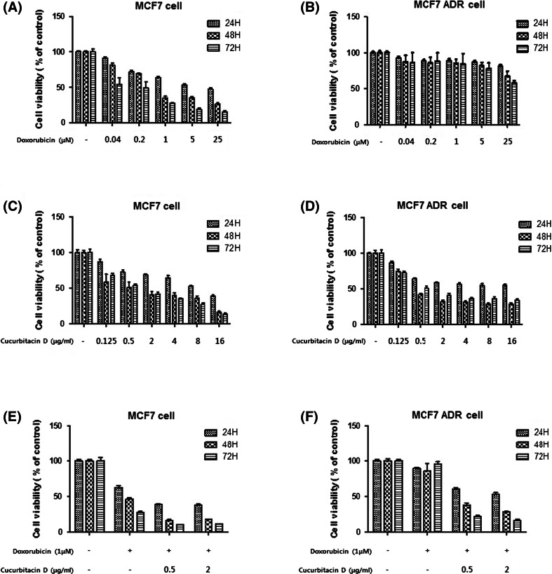
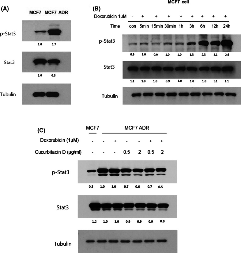
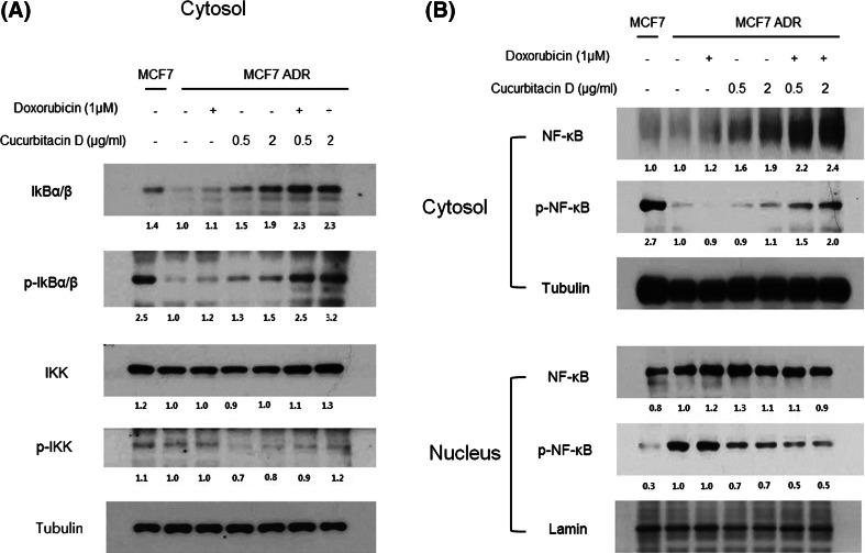
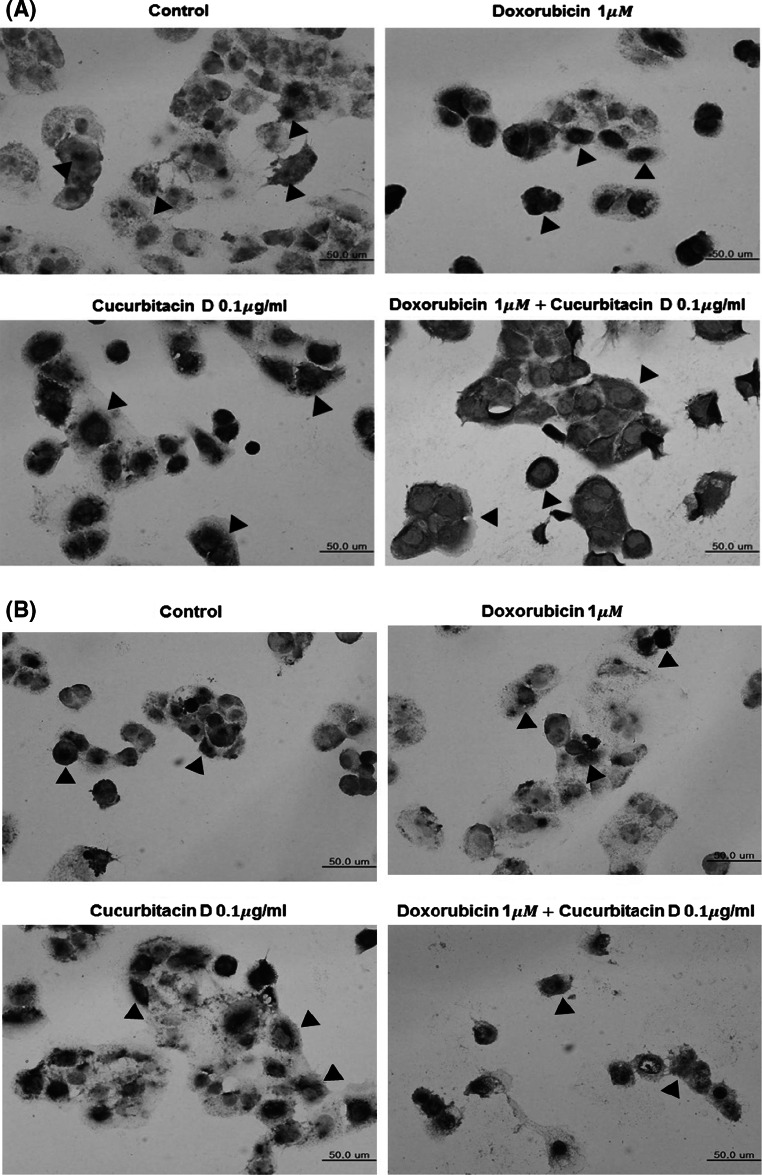
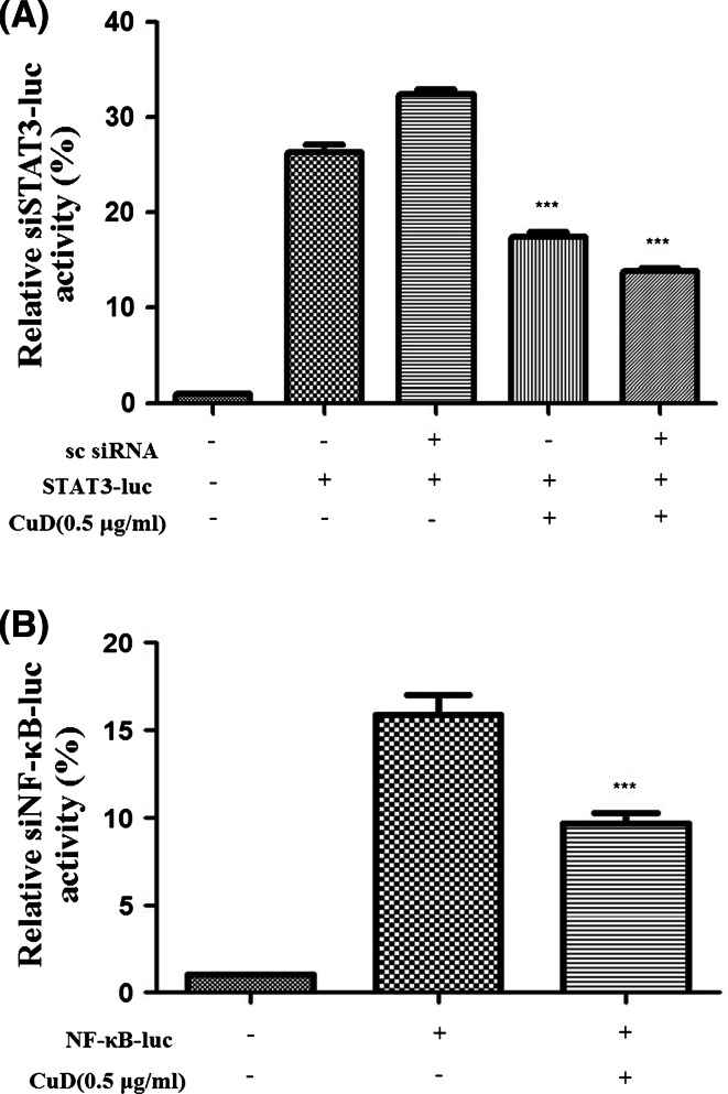
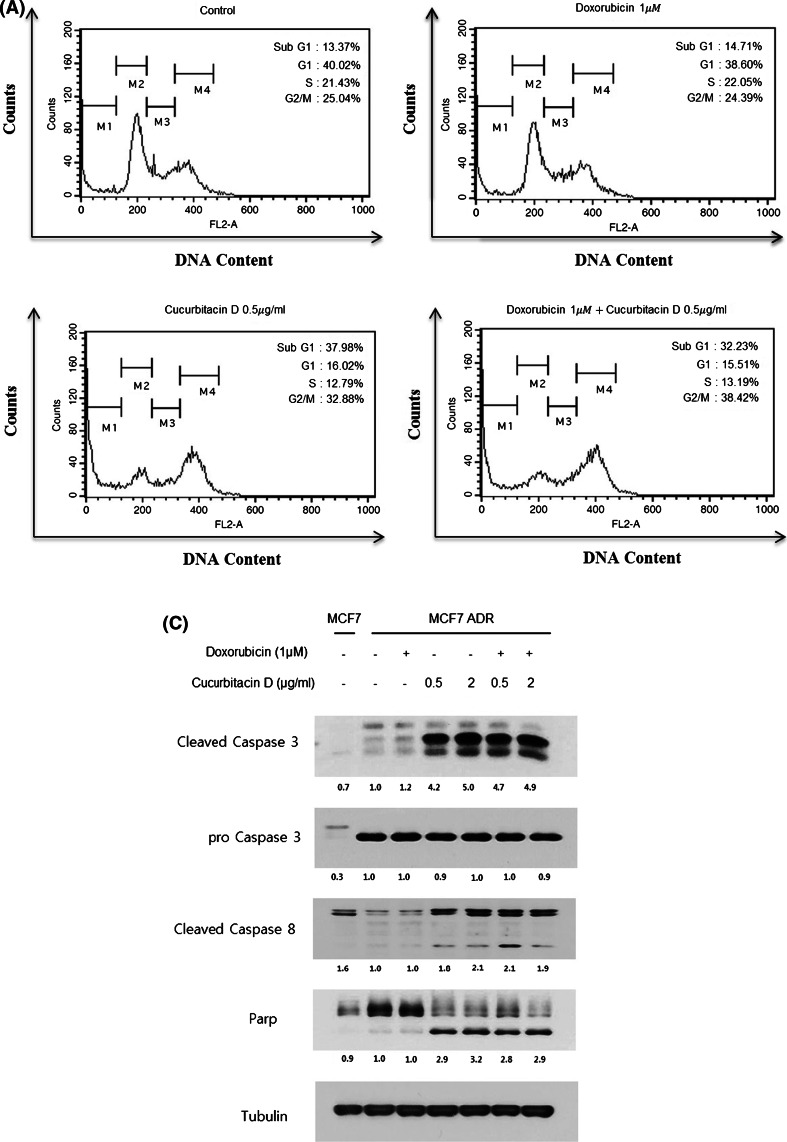
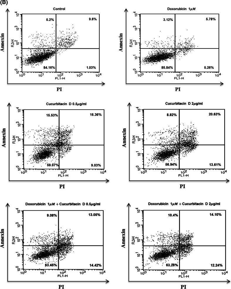
Breast cancer is the most common cancer for women and is a major cause of mortality in women. Doxorubicin is a generally used chemotherapy drug for breast cancer. However, multidrug resistance of breast cancer interferes with the chemotherapy. We examined whether cucurbitacin D affects doxorubicin resistance of MCF7/ADR breast cancer cells. Cell viability was measured by MTT assay. Levels of p-STAT3, p-NF-κB, IκB, and caspases were measured by Western blot analysis. Nuclear staining of Stat3 and NF-κB was measured by immunocytochemistry. STAT3 and NF-κB transcriptional activity was detected by STAT3 and NF-κB luciferase reporter gene assays. Analysis of cell cycle arrest was performed by flow cytometry. Induction of apoptosis by cucurbitacin D was measured by Annexin V-FITC/propidium iodide assay. More than 90% of MCF7/ADR cells lived upon treatment with doxorubicin for 24 h. However, upon treatment with cucurbitacin D, cell death was more than 60%. Co-administration of cucurbitacin D and doxorubicin induced apoptosis, and G2/M cell cycle arrest, and inhibited upregulated Stat3 by doxorubicin on MCF7/ADR cells. Additionally, cucurbitacin D led to an increase in the IκBα level in the cytosol and a decrease in the p-NF-κB level in the nucleus. Finally, cucurbitacin D inhibited translocation of Stat3 and NF-κB and decreased transcriptional activity in the nucleus. Cucurbitacin D decreases cell proliferation and induces apoptosis by inhibiting Stat3 and NF-κB signaling in doxorubicin-resistant breast cancer cells. Cucurbitacin D could be used as a useful compound to treat adriamycin-resistant patients.

Keywords: Breast cancer; Cucurbitacin D; Doxorubicin; MCF7 cell; MCF7/ADR cell; Multidrug resistance.


Figures

Fig. 1
Fig. 1
Effect of doxorubicin, cucurbitacin D, and doxorubicin with cucurbitacin D on MCF7 and MCF7/ADR cell viability. MCF7 and MCF7/ADR cells were treated with different concentrations of doxorubicin, cucurbitacin D, cucurbitacin D (0.5, 2 μg/mL), and doxorubicin (1 μM) for 24, 48, and 72 h. Cell viability was then measured using the MTT assay. Each value represents the mean ± SD. All data are p < 0.0001 by Student t-test
Fig. 2
Fig. 2
Cucurbitacin D suppresses p-Stat3 expression in MCF7/ADR Cells. Constitutive activation of Stat3 was detected in MCF7/ADR cells. p-STAT3 was strongly expressed in MCF7/ADR cells. a Doxorubicin increases constitutive STAT3 phosphorylation in a time-dependent manner in MCF7 cells. b Cucurbitacin D inhibits p-Stat3 expression in MCF7/ADR cells. c Whole-cell lysates were analyzed by Western blot with anti-pStat3, anti-Stat3, and anti-tubulin antibodies
Fig. 3
Fig. 3
Cucurbitacin D inhibits NF-κB signaling in MCF7/ADR cells. MCF7/ADR cells were treated with cucurbitacin D (0.5, 2 μg/mL) in the presence and absence of doxorubicin (1 μM). Nuclear and cytosolic extracts of cultured cells were then prepared and analyzed by Western blot to measure NF-κB levels
Fig. 4
Fig. 4
Cucurbitacin D inhibits nuclear translocation of Stat3 and NF-κB in MCF7/ADR cells. MCF7/ADR cells were treated with cucurbitacin D (0.1 μg/mL) in the presence and absence of doxorubicin (1 μM), and then submitted to immunocytochemistry for the detection of nuclear NF-κB (a) and Stat3 (b)
Fig. 5
Fig. 5
Cucurbitacin D inhibits Stat3 and NF-κB transcription in MCF7 cells. MCF7 cells were transfected with the indicated siRNA or plasmid, and then treated with each drug for 24 h. MCF7 cells were then treated with cucurbitacin D (0.5 μg/mL). Afterward, lysates were analyzed using the dual-luciferase reporter assay. Each value represents the mean ± SD. All data are p < 0.0001 by Student t-test
Fig. 6
Fig. 6
Cucurbitacin D induces G2/M cell cycle arrest and apoptosis in MCF7/ADR cells. MCF7/ADR cells were treated with cucurbitacin D (0.5 μg/mL) in the presence and absence of doxorubicin (1 μM) for 24 h. Cell cycle distribution was analyzed using a FACS flow cytometer (a). MCF7/ADR cells were treated with the indicated drugs for 24 h and subjected to Annexin V/PI assay. b Effect of cucurbitacin D on the expression of cleaved caspase-3, cleaved caspase-8, and cleaved PARP. MCF7/ADR cells were treated with cucurbitacin D (0.5 μg/mL) in the presence and absence of doxorubicin (1 μM) for 24 h. The cell lysates were subjected to Western blot analysis using specific antibodies (c)
Fig. 6
Fig. 6
Cucurbitacin D induces G2/M cell cycle arrest and apoptosis in MCF7/ADR cells. MCF7/ADR cells were treated with cucurbitacin D (0.5 μg/mL) in the presence and absence of doxorubicin (1 μM) for 24 h. Cell cycle distribution was analyzed using a FACS flow cytometer (a). MCF7/ADR cells were treated with the indicated drugs for 24 h and subjected to Annexin V/PI assay. b Effect of cucurbitacin D on the expression of cleaved caspase-3, cleaved caspase-8, and cleaved PARP. MCF7/ADR cells were treated with cucurbitacin D (0.5 μg/mL) in the presence and absence of doxorubicin (1 μM) for 24 h. The cell lysates were subjected to Western blot analysis using specific antibodies (c)

Similar articles

Cited by

References

    1. Parkin DM, Bray F, Ferlay J, Pisani P. Global cancer statistics, 2002. CA Cancer J Clin. 2005;55:74–108. doi: 10.3322/canjclin.55.2.74. - DOI - PubMed
    1. Sledge GW, Neuberg D, Bernardo P, Ingle JN, Martino S, Rowinsky EK, Wood WC. Phase III trial of doxorubicin, paclitaxel, and the combination of doxorubicin and paclitaxel as front-line chemotherapy for metastatic breast cancer: an intergroup trial (E1193) J Clin Oncol. 2003;21:588–592. doi: 10.1200/JCO.2003.08.013. - DOI - PubMed
    1. Rabbani A, Finn RM, Ausio J. The anthracycline antibiotics: antitumor drugs that alter chromatin structure. Bioessays. 2005;27:50–56. doi: 10.1002/bies.20160. - DOI - PubMed
    1. Szakacs G, Paterson JK, Ludwig JA, Booth-Genthe C, Gottesman MM. Targeting multidrug resistance in cancer. Nat Rev Drug Discov. 2006;5:219–234. doi: 10.1038/nrd1984. - DOI - PubMed
    1. Zheng A, Kallio A, Harkonen P. Tamoxifen-induced rapid death of MCF-7 breast cancer cells is mediated via extracellularly signal-regulated kinase signaling and can be abrogated by estrogen. Endocrinology. 2007;148:2764–2777. doi: 10.1210/en.2006-1269. - DOI - PubMed

Publication types

MeSH terms