. 2015 Jul 28;112(30):E4026-35.
doi: 10.1073/pnas.1503617112. Epub 2015 Jul 13.

Cloning, synthesis, and characterization of αO-conotoxin GeXIVA, a potent α9α10 nicotinic acetylcholine receptor antagonist

Affiliations

Cloning, synthesis, and characterization of αO-conotoxin GeXIVA, a potent α9α10 nicotinic acetylcholine receptor antagonist

Sulan Luo et al. Proc Natl Acad Sci U S A. .

Abstract

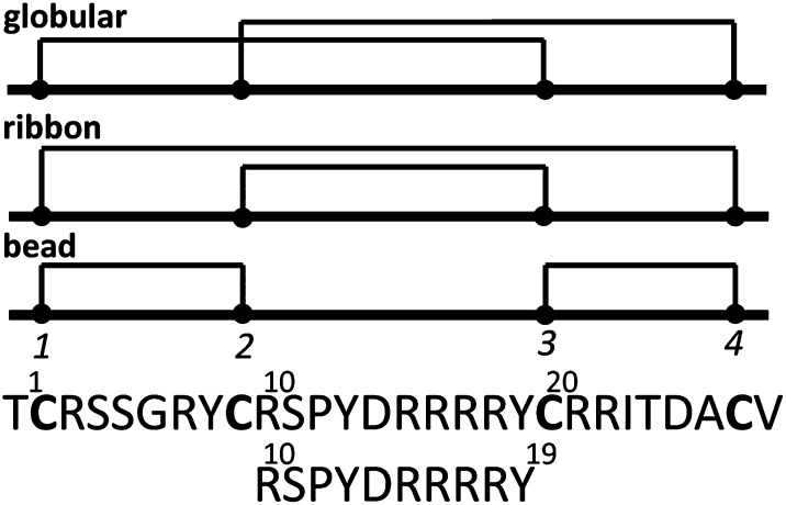
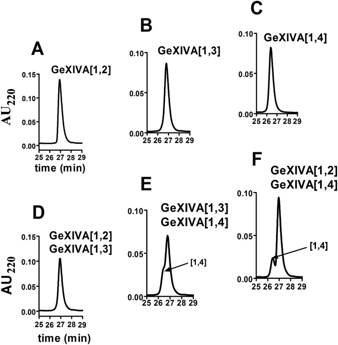
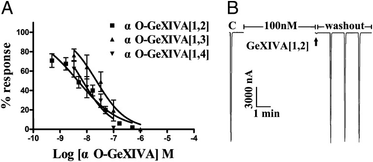
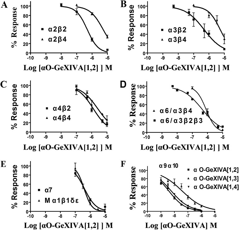
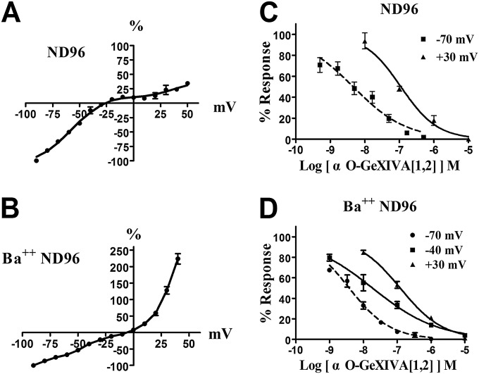
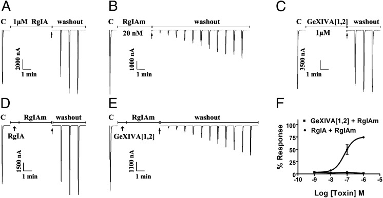
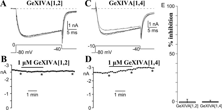
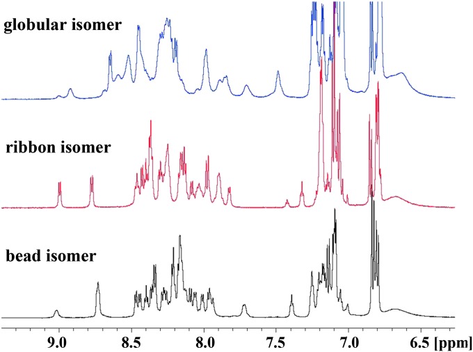
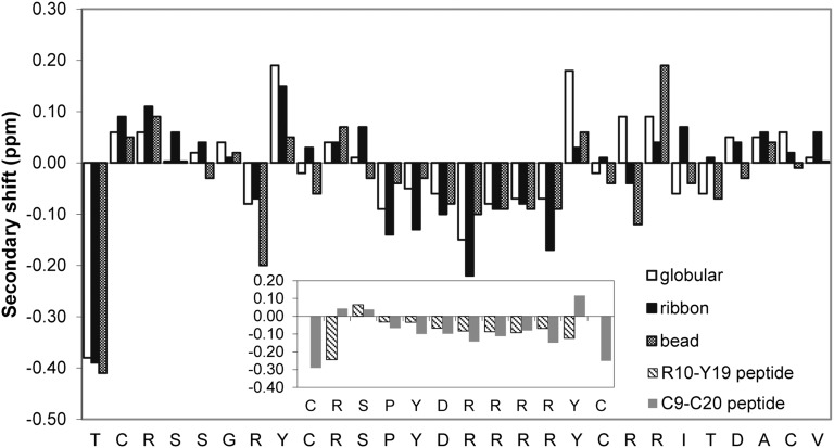
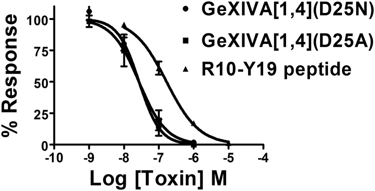
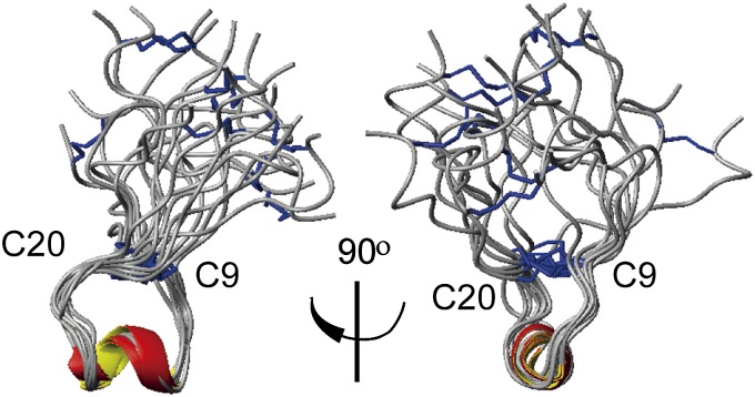
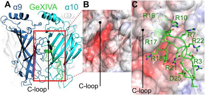
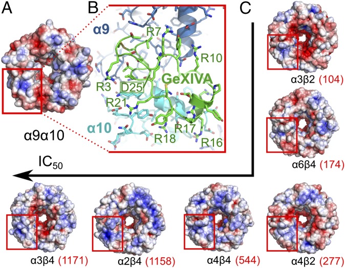
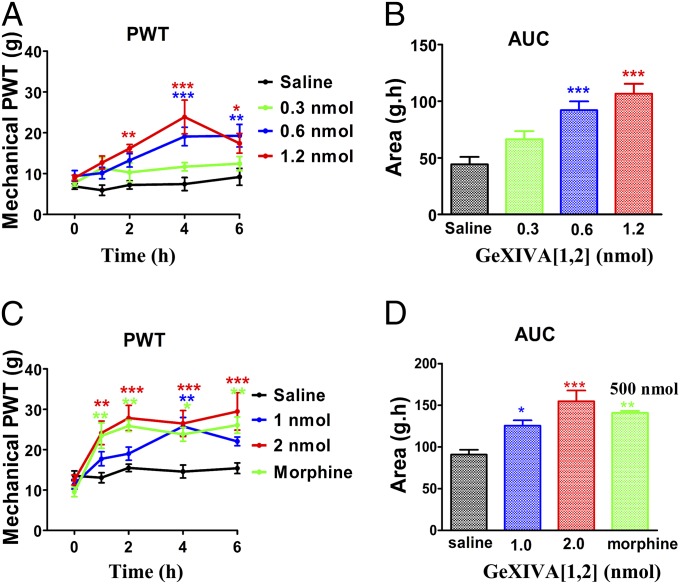
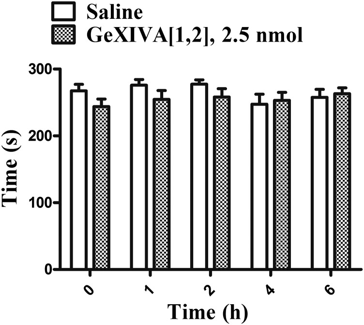
We identified a previously unidentified conotoxin gene from Conus generalis whose precursor signal sequence has high similarity to the O1-gene conotoxin superfamily. The predicted mature peptide, αO-conotoxin GeXIVA (GeXIVA), has four Cys residues, and its three disulfide isomers were synthesized. Previously pharmacologically characterized O1-superfamily peptides, exemplified by the US Food and Drug Administration-approved pain medication, ziconotide, contain six Cys residues and are calcium, sodium, or potassium channel antagonists. However, GeXIVA did not inhibit calcium channels but antagonized nicotinic AChRs (nAChRs), most potently on the α9α10 nAChR subtype (IC50 = 4.6 nM). Toxin blockade was voltage-dependent, and kinetic analysis of toxin dissociation indicated that the binding site of GeXIVA does not overlap with the binding site of the competitive antagonist α-conotoxin RgIA. Surprisingly, the most active disulfide isomer of GeXIVA is the bead isomer, comprising, according to NMR analysis, two well-resolved but uncoupled disulfide-restrained loops. The ribbon isomer is almost as potent but has a more rigid structure built around a short 310-helix. In contrast to most α-conotoxins, the globular isomer is the least potent and has a flexible, multiconformational nature. GeXIVA reduced mechanical hyperalgesia in the rat chronic constriction injury model of neuropathic pain but had no effect on motor performance, warranting its further investigation as a possible therapeutic agent.

Keywords: NMR; nicotinic; pain; α9α10 nAChR; αO-conotoxin GeXIVA.


Conflict of interest statement

Conflict of interest statement: The sequence of αO-conotoxin GeXIVA has been patented by Hainan University, with S.L., D.Z., Y.W., X.Z., Y.H., and J.M.M. listed as inventors.

Figures

Fig. 1.
Fig. 1.
Peptide sequence of αO-conotoxin GeXIVA using a one-letter code showing the disulfide connectivities of the globular, ribbon, and bead isomers in schematic form and the sequence of the fragment peptide from R10–Y19. The globular, ribbon, and bead forms are denoted throughout the main text as GeXIVA[1,3], GeXIVA[1,4], and GeXIVA[1,2], respectively, denoting the disulfide connectivity of the first disulfide bond in each case, with the second disulfide bond defined by default. Labeling of the Cys numbering is shown for the bead isomer.
Fig. S1.
Fig. S1.
Chromatographic properties of isomers of αO-conotoxin GeXIVA. HPLC chromatograms of ∼1 nmol of GeXIVA[1,2] (A); ∼1 nmol of GeXIVA[1,3] (B); ∼1 nmol of GeXIVA[1,4] (C); coelution of ∼0.33 nmol each of GeXIVA[1,2] + GeXIVA[1,3] (D); coelution of ∼0.33 nmol each of GeXIVA[1,3] + GeXIVA[1,4] (E); and coelution of ∼0.33 nmol each of GeXIVA[1,2] + GeXIVA[1,4] (F). Note that GeXIVA[1,2] + GeXIVA[1,3] coelute, necessitating synthetic preparation by directed disulfide folding. Peptides were analyzed on RP analytical Vydac C18 HPLC using a linear gradient of 10–50% (vol/vol) buffer B60 for 40 min. Buffer B60 was 60% (vol/vol) acetonitrile, 40% (vol/vol) H20, and 0.092% trifluoroacetic acid; buffer A was 0.1% trifluoroacetic acid in H2O. A at 220 nm was monitored.
Fig. 2.
Fig. 2.
αO-conotoxin GeXIVA blocks rat α9α10 nAChRs. (A) Concentration response of GeXIVA isomers on α9α10 nAChRs. Oocytes expressing α9α10 nAChR were voltage-clamped at −70 mV and subjected to a 1-s pulse of 10 μM ACh every minute as described in Materials and Methods. The IC50s were as follows: GeXIVA[1,2], 4.6 nM (3.18–6.65 nM); GeXIVA[1,3], 22.7 nM (11.8–43.5 nM); and GeXIVA[1,4], 7 nM (3.6–13.4 nM). Data points are mean ± SEM. The numbers inside the parentheses in the legend indicate 95% confidence interval of the IC50 or Hill slope. Hill slopes were GeXIVA[1,2], 0.56 (0.44–0.69); GeXIVA[1,3], 0.78 (0.29–1.26); and GeXIVA[1,4], 0.79 (0.23–1.36). Values were from six to 12 separate oocytes. (B) Representative block of ACh response of α9α10 nAChRs by 100 nM GeXIVA[1,2]. The “C” responses are control, following which the oocyte was exposed to peptide for 5 min. The arrow denotes the first ACh response in the presence of toxin. Peptide was then washed out, and subsequent responses to ACh were measured at 1-min intervals. Blockade of α9α10 nAChRs was rapidly reversed.
Fig. S2.
Fig. S2.
Concentration responses of αO-conotoxin GeXIVA[1,2] on rat nAChR subtypes (AE) and αO-conotoxin GeXIVA isomers on rat α9α10 nAChR in the presence of Ba++ (F). (AE) IC50s and Hill slopes are shown in Table 2. Note that the peptide is generally more potent on β2- vs. β4-containing nAChRs. (F) Concentration response of equimolar Ba++ was substituted for Ca2+ in the perfusion solution, as described in Materials and Methods, to prevent activation of endogenous Xenopus oocyte Ca2+-activated Cl currents. Values are mean ± SEM from six to nine separate oocytes. The IC50 for GeXIVA[1,2] was 3.8 nM (3.1–4.8 nM) with a Hill slope of 0.71 (0.58–0.84). The IC50 for GeXIVA[1,3] was 37 nM (25.0–55.7 nM) with a Hill slope of 0.54 (0.42–0.65). The IC50 for GeXIVA[1,4] was 5.8 nM (4.7–7.1 nM) with a Hill slope of 0.65 (0.56–0.73). The numbers inside the parentheses in the legend indicate 95% confidence interval of the IC50 or Hill slope.
Fig. S3.
Fig. S3.
Voltage dependence of α9α10 nAChR blockade by αO-conotoxin GeXIVA[1,2]. Current responses were measured at membrane-holding potentials that were varied in 10-mV steps once per minute. Current–voltage responses were obtained in ND96 buffer (A) and buffer in which Ca2+ was replaced by Ba++ (Ba++ ND96) (B). Values were normalized to the response measured at −90 mV and represent the mean ± SEM. Concentration responses of αO-GeXIVA[1,2] on rat α9α10 nAChR at −70 mV and +30 mV in ND-96 (C) and at −70 mV, −40 mV, and +30 mV in Ba++ ND-96 (D) are shown. Values are mean ± SEM from five to 12 separate oocytes. In C, the IC50s in ND96 were −70 mV, 4.6 nM (3.2–6.7 nM) and +30 mV, 111 nM (73.8–166 nM). Hill slopes in ND96 were −70 mV, 0.56 (0.44–0.69) and +30 mV, 0.82 (0.56–1.08). In D, the IC50s in Ba++ ND96 were −70 mV, 3.8 nM (3.1–4.8 nM); −40 mV, 18.7 nM (1.7–29.9 nM); and +30 mV, 123 nM (100–151 nM). Hill slopes in Ba++ ND96 were −70 mV, 0.71 (0.58–0.84); −40 mV, 0.45 (0.35–0.55); and +30 mV, 0.68 (0.59–0.78). The numbers inside the parentheses in the legend indicate 95% confidence interval of the IC50 or Hill slope. Values are mean ± SEM.
Fig. 3.
Fig. 3.
Binding site comparison of αO-GeXIVA and α-conotoxin RgIAm. In each instance, toxin solution was applied to α9α10 nAChRs expressed in Xenopus oocytes, followed by toxin washout. The response to 1-s pulses to ACh were measured as described in Materials and Methods. (A) One micromolar α-conotoxin RgIA for 5 min. Note the rapid recovery following toxin washout. (B) Twenty nanomolar α-conotoxin RgIAm for 4 min. Note the slow recovery. (C) One micromolar αO-conotoxin GeXIVA[1,2] for 5 min. Note the rapid recovery. (D) One micromolar α-conotoxin RgIA for 1 min, followed by 20 nM α-conotoxin RgIAm for 4 min. Note that preblockade of α9α10 nAChR with α-conotoxin RgIA prevented the slowly reversible block associated with α-conotoxin RgIAm, consistent with competitive binding to the same or overlapping site. (E) One micromolar αO-GeXIVA[1,2] for 1 min, followed by 20 nM α-conotoxin RgIAm for 4 min. Note that preblockade of α9α10 nAChR with αO-GeXIVA[1,2] did not prevent the very slowly reversible block associated with α-conotoxin RgIAm, consistent with occupancy of distinct binding sites. (F) Oocytes expressing α9α10 nAChRs were preincubated with varying concentrations of RgIA for 5 min. This step was followed by coincubation with RgIA + 20 nM RgIAm for a further 5 min. Both toxins were then washed out, and the response was measured following 2 min of washout. Note that an increasing concentration of RgIA led to a greater percent response (faster recovery) to ACh. Oocytes expressing α9α10 nAChRs were preincubated with varying concentrations of GeXIVA[1,2] for 5 min. This step was followed by coincubation with GeXIVA[1,2] + 20 nM RgIAm for a further 5 min. Both toxins were then washed out, and the response was measured following 2 min of washout. Note that an increasing concentration of GeXIVA[1,2] had no effect on percent response to ACh (n = 5–6 oocytes).
Fig. S4.
Fig. S4.
αO-contoxins GeXIVA[1,2] and GeXIVA[1,4] do not affect voltage-dependent calcium current in rat DRG neurons. Different DRG neurons were exposed for 2.5 min to 1 μM of either GeXIVA[1,2] (A) or GeXIVA[1,4] (C). Currents were elicited at the indicated voltages (Materials and Methods) once every 10 s, and are shown before (gray trace), during (black trace), and upon (gray trace) recovery from toxin. The charge carrier was 5 mM Ba2+. (B and D) Time course is shown for the data depicted above. The asterisks indicate the times when the currents shown above were generated. The solid bar indicates the duration of toxin application. (E) Average effect of toxin on voltage-dependent calcium current (n = 8, 8). The duration of toxin application ranged from 1.2 to 3.1 min for GeXIVA[1,2] (mean = 1.7 min) and from 1.5 to 2.5 min for GeXIVA[1,4] (mean = 1.9 min). Neither toxin significantly affected calcium current (P > 0.05). The neuronal diameter ranged from 21 to 34 μm (mean = 27 μm) for the GeXIVA[1,2]-exposed cells and from 26 to 32 μm (mean = 30 μm) for the GeXIVA[1,4]-exposed cells.
Fig. 4.
Fig. 4.
Amide region of the 1H-NMR spectra of GeXIVA recorded at 298 K and 600 MHz. Globular, GeXIVA[1,3]; ribbon, GeXIVA[1,4]; and bead, GeXIVA[1,2] isomers are shown.
Fig. S5.
Fig. S5.
Secondary αH shifts of the three isomers of GeXIVA (globular, ribbon, and bead) in water. (Inset) Secondary αH shifts of synthetic peptides corresponding to residues from R10–Y19 and from C9–C20.
Fig. S6.
Fig. S6.
Concentration response curves of GeXIVA analogs. Compounds were tested on α9α10 nAChRs expressed in Xenopus oocytes. The peptide with sequence RSPYDRRRY had an IC50 of 168 (129–217) nM, with a Hill slope of 0.93 (0.71–1.1). GeXIVA[1,4](D25N) had an IC50 of 27 (18–42) nM, with a Hill slope of 1.4 (0.89–2.0). GeXIVA[1,4](D25A) had an IC50 of 27 (15–50) nM, with a Hill slope of 1.1 (0.53–1.7). The numbers inside the parentheses in the legend indicate 95% confidence interval of the IC50 or Hill slope. Values are mean ± SEM (n = 3–6 oocytes per data point).
Fig. 5.
Fig. 5.
Ribbon diagram of the NMR solution structure of GeXIVA[1,4]. Images are overlaid across the C9–C20 loop. Disulfide bonds are depicted in blue. α-Helical regions are colored red and yellow.
Fig. 6.
Fig. 6.
Model of the interaction between GeXIVA[1,4] and the α9α10 subunit orthosteric binding site located between α9 (principal) and α10 (complementary) subunits. (A) Binding site is mainly contributed by the C-loop of the α9 subunit and a β-sheet from the α10 subunit. The α-helix of GeXIVA binds at the same location as framework I α-conotoxin but in a reverse orientation. (B) Electrostatic potential generated by the α9α10 ligand-binding domain mapped on the solvent-accessible surface of the GeXIVA-binding site. The solvent-accessible surface is colored from red to blue corresponding to electrostatic potentials from −5 kT/e and below to +5 kT/e and above, respectively. The binding site creates a negative charge potential compatible with the binding of GeXIVA, which is highly positively charged. (C) GeXIVA is tightly packed in the binding site, and GeXIVA-charged side chains R16 and R17 establish salt bridges deep in the pocket in the model. The molecular model was built by homology with the crystal structure of AChBP in complex with conotoxin [A10L,D14k]PnIA [Protein Data Bank (PDB) ID code 2BR8], as well as the extracellular domain of the monomer α1 subunit (PDB ID code 2QC1). The model was refined by 15-ns molecular dynamics simulations.
Fig. 7.
Fig. 7.
Potential second binding site of GeXIVA [1,4] on the extracellular domain of α9α10 and other nAChRs. (A) α9α10 Subtype displays an electropositive pocket surrounded by two negatively charged patches next to the central pore on the ligand-binding domain. (B) This patch was identified by coarse-grained molecular docking as a potential interaction site of GeXIVA, with its side chain, D25, occupying the positively charged pocket and several of its Arg side chains establishing charges and salt bridge interactions with positively charged residues of α9 and α10 subunits. (C) Same motif of electrostatic potential could be identified on the surface of other tested nAChR subtypes, and the integrity of this motif seems to correlate with the activity of GeXIVA. The fold difference from the activity on α9α10 is given in parentheses. The molecular models of the nAChR ligand-binding domains were built by homology with AChBP structure (PDB ID code 2BR8), the extracellular domain of monomer α1 subunit (PDB ID code 2QC1), and the structure of the β-subunit in the EM structure of Torpedo marmorata muscle type nAChR (PDB ID code 2BG9). The solvent-accessible surfaces were colored from red to blue, corresponding to electrostatic potentials from −5 kT/e and below to +5 kT/e and above, respectively.
Fig. 8.
Fig. 8.
Effect of αO-conotoxin GeXIVA[1,2] on mechanical hyperalgesia. The sciatic nerve of rats was loosely ligated to produce CCI, a model of human neuropathic pain, as described in Materials and Methods. Rats were injected i.m. with 0.9% saline, conotoxin, or morphine. PWT was used as a measure of mechanical hyperalgesia. (A) Effect of compound vs. time was assessed using a series of von Frey hairs. Time point t = 0 represents mechanical PWT immediately before injection. (B) Corresponding dose–response calculated as AUC for data from each dose of drug in A for time points between 0 and 6 h. (C) PWT was assessed for a separate group of rats using an electronic von Frey anesthesia meter. (D) AUC for data from each dose of drug in C. (n = 8 for A and B; n = 6 for C and D.) Each point in A and C represents the mean ± SEM. *P < 0.05, **P < 0.01, ***P < 0.001; Dunnett’s post hoc tests.
Fig. S7.
Fig. S7.
Effect of the peptide αO-conotoxin GeXIVA[1,2] on motor performance. Each bar represents the mean ± SEM change in rotarod latency (s) of preinjection (t = 0) and at 1 h, 2 h, 4 h, and 6 h after injection with saline or peptide (2.5 nmol) (n = 8).

References

    1. Olivera BM, Teichert RW. Diversity of the neurotoxic Conus peptides: A model for concerted pharmacological discovery. Mol Interv. 2007;7(5):251–260. - PubMed
    1. Mayer AM, et al. The odyssey of marine pharmaceuticals: A current pipeline perspective. Trends Pharmacol Sci. 2010;31(6):255–265. - PubMed
    1. Kaas Q, Westermann JC, Craik DJ. Conopeptide characterization and classifications: An analysis using ConoServer. Toxicon. 2010;55(8):1491–1509. - PubMed
    1. Gotti C, et al. Structural and functional diversity of native brain neuronal nicotinic receptors. Biochem Pharmacol. 2009;78(7):703–711. - PubMed
    1. Beech RN, Callanan MK, Rao VT, Dawe GB, Forrester SG. Characterization of cys-loop receptor genes involved in inhibitory amine neurotransmission in parasitic and free living nematodes. Parasitol Int. 2013;62(6):599–605. - PubMed

Publication types

MeSH terms

Associated data