

Partial hepatectomy induces delayed hepatocyte proliferation and normal liver regeneration in ovariectomized mice
- PMID: 26170710
- PMCID: PMC4494181
- DOI: 10.2147/CEG.S80212
Partial hepatectomy induces delayed hepatocyte proliferation and normal liver regeneration in ovariectomized mice
Abstract
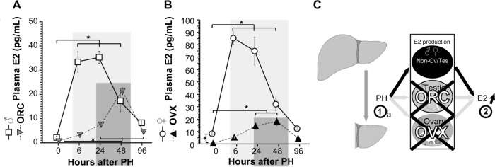
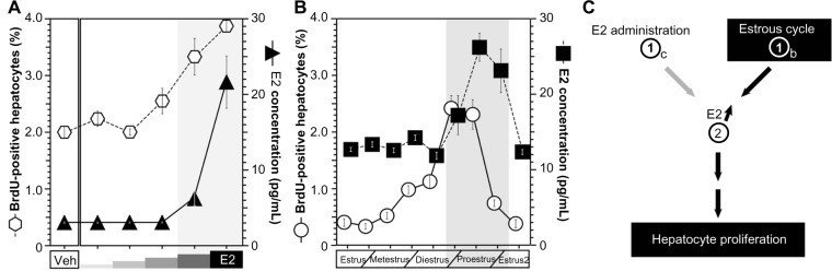
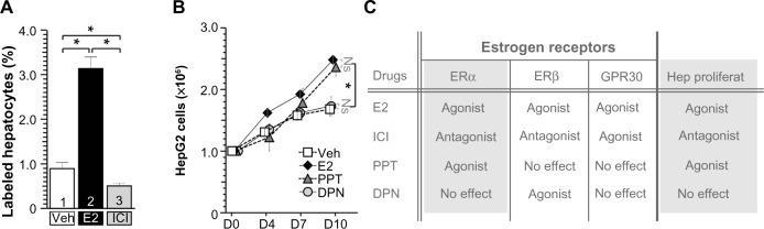
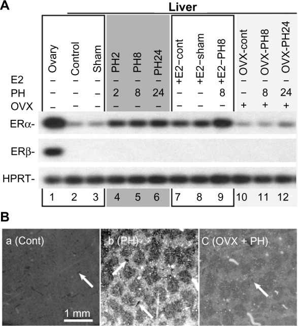
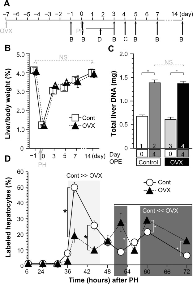
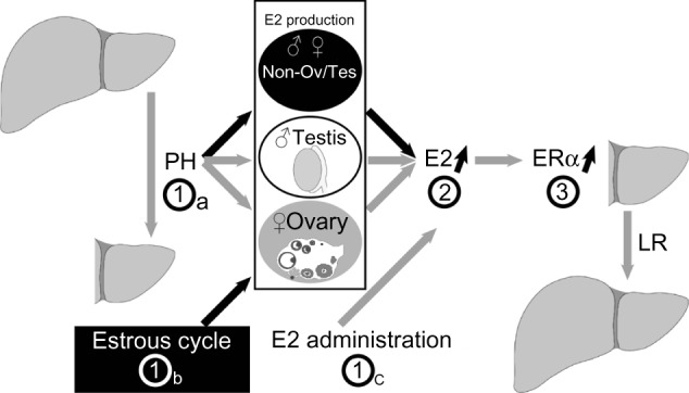
Estrogens play central roles in sexual development, reproduction, and hepatocyte proliferation. The ovaries are one of the main organs for estradiol (E2) production. Ovariectomies (OVXs) were performed on the female mice, and hepatocyte proliferation was analyzed. The ovariectomized mice exhibited delayed hepatocyte proliferation after partial hepatectomy (PH) and also exhibited delayed and reduced E2 induction. Both E2 administration and PH induced the gene expression of estrogen receptor α (ERα). The transcripts of ERα were detected specifically in periportal hepatocytes after E2 administration and PH. Moreover, the E2 concentrations and hepatocyte proliferation rates were highest in the proestrus period of the estrous cycle. Taken together, these findings indicate that E2 accelerated ERα expression in periportal hepatocytes and hepatocyte proliferation in the female mice.
Keywords: ER; estrogen; estrous cycle; hepatocyte proliferation; liver regeneration.
Figures
Similar articles
-
Estradiol accelerates liver regeneration through estrogen receptor α.Clin Exp Gastroenterol. 2019 Jul 22;12:331-336. doi: 10.2147/CEG.S214196. eCollection 2019. Clin Exp Gastroenterol. 2019. PMID: 31413616 Free PMC article.
-
Estrogen induces estrogen receptor alpha expression and hepatocyte proliferation in the livers of male mice.Genes Cells. 2015 Mar;20(3):217-23. doi: 10.1111/gtc.12214. Epub 2014 Dec 12. Genes Cells. 2015. PMID: 25495062
-
Estrogen induces estrogen receptor α expression and hepatocyte proliferation in late pregnancy.Biochem Biophys Res Commun. 2019 Apr 9;511(3):592-596. doi: 10.1016/j.bbrc.2019.02.119. Epub 2019 Feb 28. Biochem Biophys Res Commun. 2019. PMID: 30826053
-
Effects of prolactin and estrogen on cell proliferation of the mouse liver induced by partial hepatectomy.In Vivo. 1997 Sep-Oct;11(5):409-13. In Vivo. 1997. PMID: 9427045
-
Estrogen Accelerates Cell Proliferation through Estrogen Receptor α during Rat Liver Regeneration after Partial Hepatectomy.Acta Histochem Cytochem. 2017 Feb 28;50(1):39-48. doi: 10.1267/ahc.17003. Epub 2017 Feb 25. Acta Histochem Cytochem. 2017. PMID: 28386149 Free PMC article.
Cited by
-
UDP-glucose, cereblon-dependent proinsulin degrader.Sci Rep. 2022 Aug 26;12(1):14568. doi: 10.1038/s41598-022-18902-5. Sci Rep. 2022. PMID: 36028536 Free PMC article.
-
Nuclear ErbB2 expression in hepatocytes in liver disease.Virchows Arch. 2021 Feb;478(2):309-318. doi: 10.1007/s00428-020-02871-z. Epub 2020 Jun 26. Virchows Arch. 2021. PMID: 32591879 Free PMC article.
-
Chorionic gonadotropin stimulates maternal hepatocyte proliferation during pregnancy.Biochem Biophys Res Commun. 2021 Nov 19;579:110-115. doi: 10.1016/j.bbrc.2021.09.039. Epub 2021 Sep 24. Biochem Biophys Res Commun. 2021. PMID: 34597993 Free PMC article.
-
Estrogen Activation of G-Protein-Coupled Estrogen Receptor 1 Regulates Phosphoinositide 3-Kinase and mTOR Signaling to Promote Liver Growth in Zebrafish and Proliferation of Human Hepatocytes.Gastroenterology. 2019 May;156(6):1788-1804.e13. doi: 10.1053/j.gastro.2019.01.010. Epub 2019 Jan 12. Gastroenterology. 2019. PMID: 30641053 Free PMC article.
-
Estradiol accelerates liver regeneration through estrogen receptor α.Clin Exp Gastroenterol. 2019 Jul 22;12:331-336. doi: 10.2147/CEG.S214196. eCollection 2019. Clin Exp Gastroenterol. 2019. PMID: 31413616 Free PMC article.
References
LinkOut - more resources
Full Text Sources
Other Literature Sources
