

Nonadherent culture method downregulates stem cell antigen-1 expression in mouse bone marrow mesenchymal stem cells
- PMID: 26170908
- PMCID: PMC4486893
- DOI: 10.3892/etm.2015.2457
Nonadherent culture method downregulates stem cell antigen-1 expression in mouse bone marrow mesenchymal stem cells
Abstract
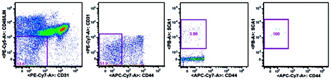
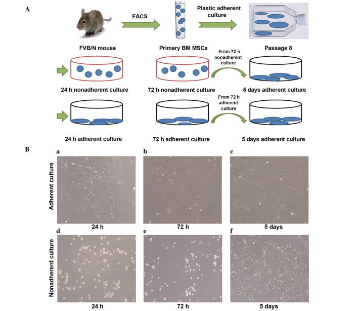
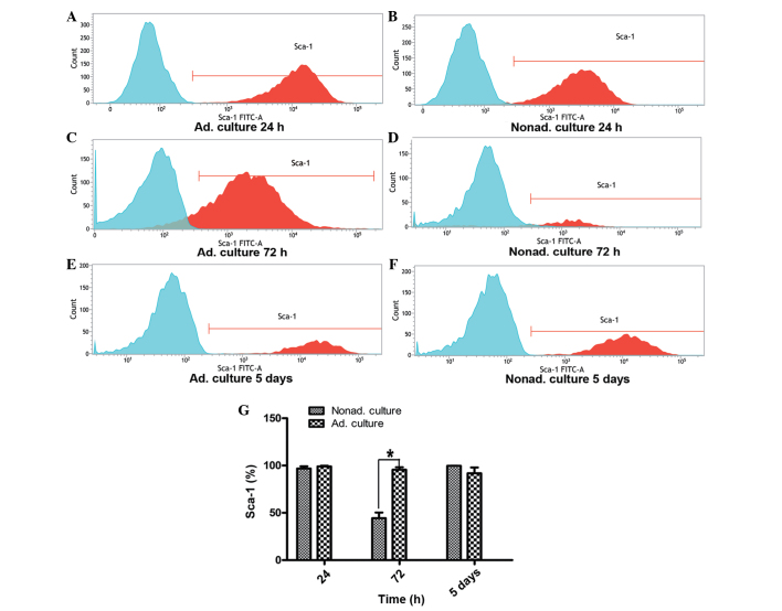
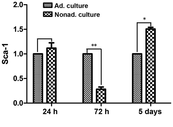
Mesenchymal stem cells (MSCs) are primarily isolated by their adherence to plastic and their in vitro growth characteristics. Expansion of these cells from an adherent culture is the only method to obtain a sufficient number of cells for use in clinical practice and research. However, little is known with regard to the effect of adherence to plastic on the phenotype of the cells. In the present study, bone marrow CD45-CD31-CD44- stem cell antigen (Sca)-1+ MSCs were sorted by flow cytometry and expanded in adherent cultures. The expression levels of the adhesion molecule, Sca-1, in the adherent cultures were compared with those from nonadherent cultures at different time points. The flow cytometry results indicated that the expression levels of Sca-1 decreased in the MSCs in the nonadherent cultures grown in ultra-low-adherent plates. Furthermore, the result was confirmed by quantitative polymerase chain reaction at the same time points. Therefore, the results demonstrated that the loss of plastic adherence downregulated the expression of Sca-1. The observations may provide novel insights into the molecular mechanisms underlying plastic adherent culture.
Keywords: adherent culture; mesenchymal stem cells; nonadherent culture; phenotype; stem cell antigen-1.
Figures
Similar articles
-
Removal from adherent culture contributes to apoptosis in human bone marrow mesenchymal stem cells.Mol Med Rep. 2017 Jun;15(6):3499-3506. doi: 10.3892/mmr.2017.6440. Epub 2017 Apr 7. Mol Med Rep. 2017. PMID: 28393226 Free PMC article.
-
An efficient method for isolation of murine bone marrow mesenchymal stem cells.Int J Dev Biol. 2007;51(8):723-9. doi: 10.1387/ijdb.072352ns. Int J Dev Biol. 2007. PMID: 17939119
-
Isolation murine mesenchymal stem cells by positive selection.In Vitro Cell Dev Biol Anim. 2007 Sep-Oct;43(8-9):276-82. doi: 10.1007/s11626-007-9041-5. Epub 2007 Sep 12. In Vitro Cell Dev Biol Anim. 2007. PMID: 17851725
-
Characterization of mesenchymal stem cells isolated from murine bone marrow by negative selection.J Cell Biochem. 2003 Aug 15;89(6):1235-49. doi: 10.1002/jcb.10594. J Cell Biochem. 2003. PMID: 12898521
-
Flow cytometric discrimination of mesenchymal progenitor cells from bone marrow-adherent cell populations using CD34/44/45(-) and Sca-1(+) markers.J Orthop Sci. 2007 Mar;12(2):161-9. doi: 10.1007/s00776-006-1098-6. Epub 2007 Mar 30. J Orthop Sci. 2007. PMID: 17393272
Cited by
-
Nonadherent culture method promotes MSC-mediated vascularization in myocardial infarction via miR-519d/VEGFA pathway.Stem Cell Res Ther. 2020 Jul 2;11(1):266. doi: 10.1186/s13287-020-01780-x. Stem Cell Res Ther. 2020. PMID: 32616068 Free PMC article.
-
Removal from adherent culture contributes to apoptosis in human bone marrow mesenchymal stem cells.Mol Med Rep. 2017 Jun;15(6):3499-3506. doi: 10.3892/mmr.2017.6440. Epub 2017 Apr 7. Mol Med Rep. 2017. PMID: 28393226 Free PMC article.
-
Identification of Stromal Cells in Spleen Which Support Myelopoiesis.Front Cell Dev Biol. 2019 Jan 24;7:1. doi: 10.3389/fcell.2019.00001. eCollection 2019. Front Cell Dev Biol. 2019. PMID: 30733944 Free PMC article.
References
LinkOut - more resources
Full Text Sources
Other Literature Sources
Research Materials
Miscellaneous