Primary glioblastoma of the cerebellar vermis: A case report
- PMID: 26171039
- PMCID: PMC4487146
- DOI: 10.3892/ol.2015.3188
Primary glioblastoma of the cerebellar vermis: A case report
Abstract
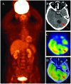
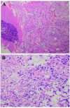
Cerebellar glioblastoma is a rare adult tumor. The accurate diagnosis of cerebellar glioblastoma is important for establishing a suitable therapeutic schedule. However, it is occasionally difficult to diagnosis these tumors. Clinical presentation, computed tomography (CT) and magnetic resonance imaging can provide useful information, but they may not lead to a definitive diagnosis. Positron emission tomography/computed tomography (PET/CT) may provide a novel way of forming a differential diagnoses. The lesions of glioblastoma multiforme (GBM) rarely occur in the cerebellum, with prior studies reporting that only 0.4-3.4% of all GBM tumors occur here. In the current study, a case of primary cerebellar glioblastoma is presented and the physiopathology, clinical presentation, diagnosis, differential diagnosis, treatment and general outcome of this disease are discussed. A 61-year-old female presented with nausea, vomiting, balance problems and cerebellar signs. Cranial magnetic resonance imaging (MRI) and positron emission tomography/computed tomography (PET/CT) examination demonstrated one regular contour of a mass lesion in the cerebellar vermis. Following surgery, glioblastoma was histologically confirmed. The outcome of the patient was favorable after 18 months of follow-up. Cerebellar GBM should be considered in the differential diagnosis of a cerebellar mass lesion, and PET/CT may provide a novel identification method for different cerebellar mass lesions.
Keywords: cerebellar; fluorodeoxyglucose; glioblastoma; magnetic resonance imaging; positron emission tomography/computed tomography.
Figures


Similar articles
-
Primary glioblastoma of the cerebellum in a 19-year-old woman: a case report.J Med Case Rep. 2012 Oct 2;6:329. doi: 10.1186/1752-1947-6-329. J Med Case Rep. 2012. PMID: 23031548 Free PMC article.
-
Radiological features of cerebellar glioblastoma.J Neuroradiol. 2016 Jul;43(4):260-5. doi: 10.1016/j.neurad.2015.10.006. Epub 2015 Dec 28. J Neuroradiol. 2016. PMID: 26740386
-
Glioblastoma multiforme of the cerebellum in an elderly man.J Chin Med Assoc. 2004 Jun;67(6):301-4. J Chin Med Assoc. 2004. PMID: 15366408
-
[Glioblastoma multiforme developing separately from the initial lesion 9 years after successful treatment for gliomatosis cerebri: a case report].No Shinkei Geka. 2008 Aug;36(8):709-15. No Shinkei Geka. 2008. PMID: 18700534 Review. Japanese.
-
Cerebellar glioblastoma multiforme in an adult.Arq Neuropsiquiatr. 2006 Mar;64(1):132-5. doi: 10.1590/s0004-282x2006000100028. Epub 2006 Apr 5. Arq Neuropsiquiatr. 2006. PMID: 16622570 Review.
Cited by
-
Characteristics of cerebellar glioblastomas in adults.J Neurooncol. 2018 Feb;136(3):555-563. doi: 10.1007/s11060-017-2682-7. Epub 2017 Dec 1. J Neurooncol. 2018. PMID: 29196927
-
Cerebellar cystic glioblastomas: An uncommon presentation of a rare disease and clinical review.eNeurologicalSci. 2018 Dec 17;14:60-61. doi: 10.1016/j.ensci.2018.12.002. eCollection 2019 Mar. eNeurologicalSci. 2018. PMID: 30623119 Free PMC article. No abstract available.
References
LinkOut - more resources
Full Text Sources
Other Literature Sources
