

Computed Tomography Staging of Middle Ear Cholesteatoma
- PMID: 26171086
- PMCID: PMC4485650
- DOI: 10.12659/PJR.894155
Computed Tomography Staging of Middle Ear Cholesteatoma
Abstract
Background: To establish computed tomography (CT) staging of middle ear cholesteatoma and assess its impact on the selection of the surgical procedure.
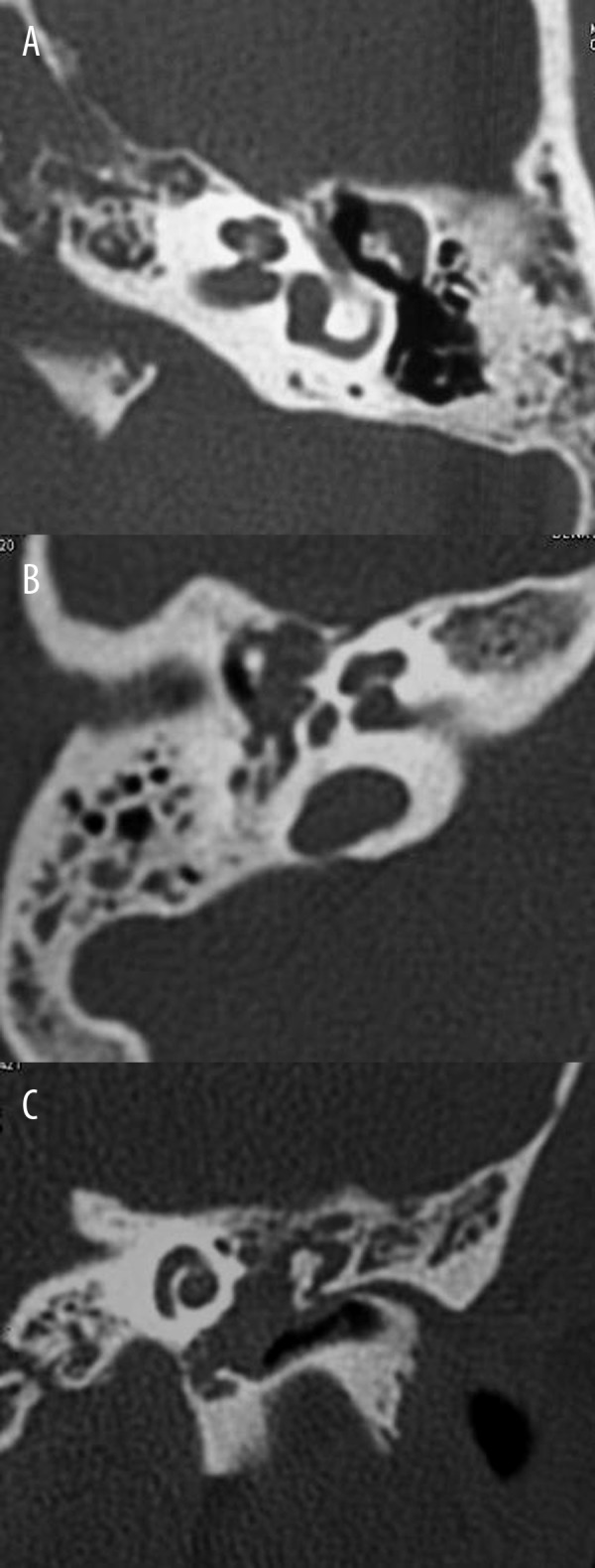
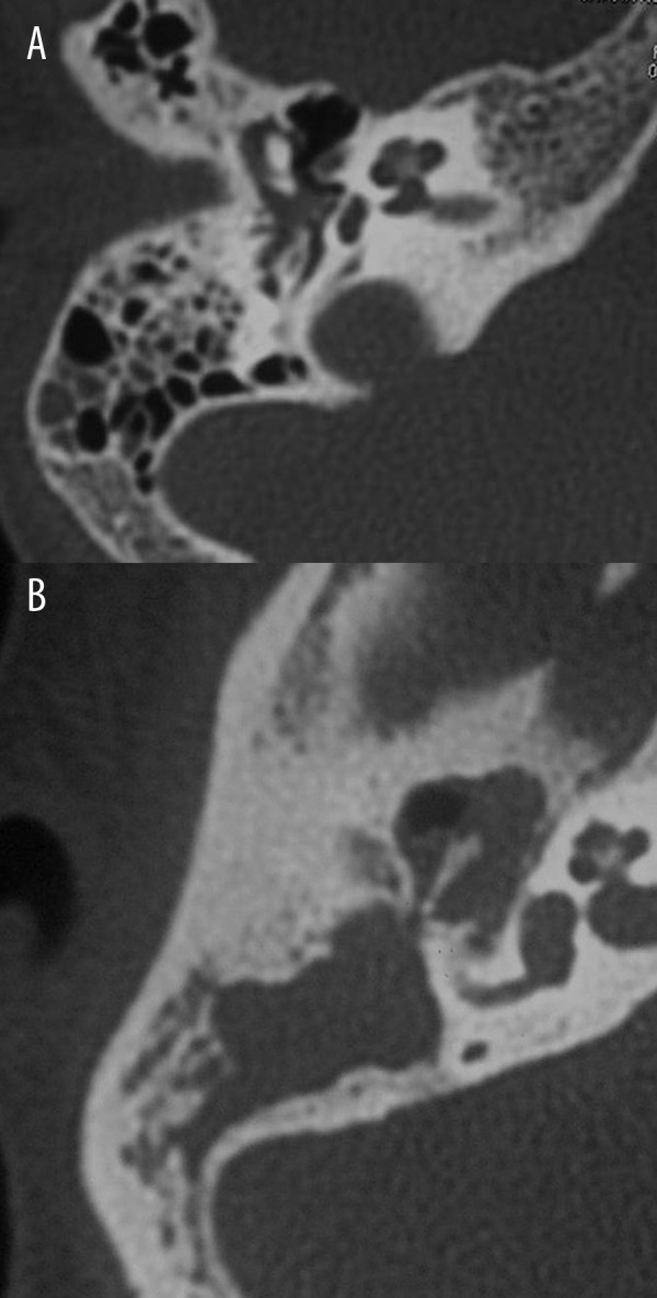
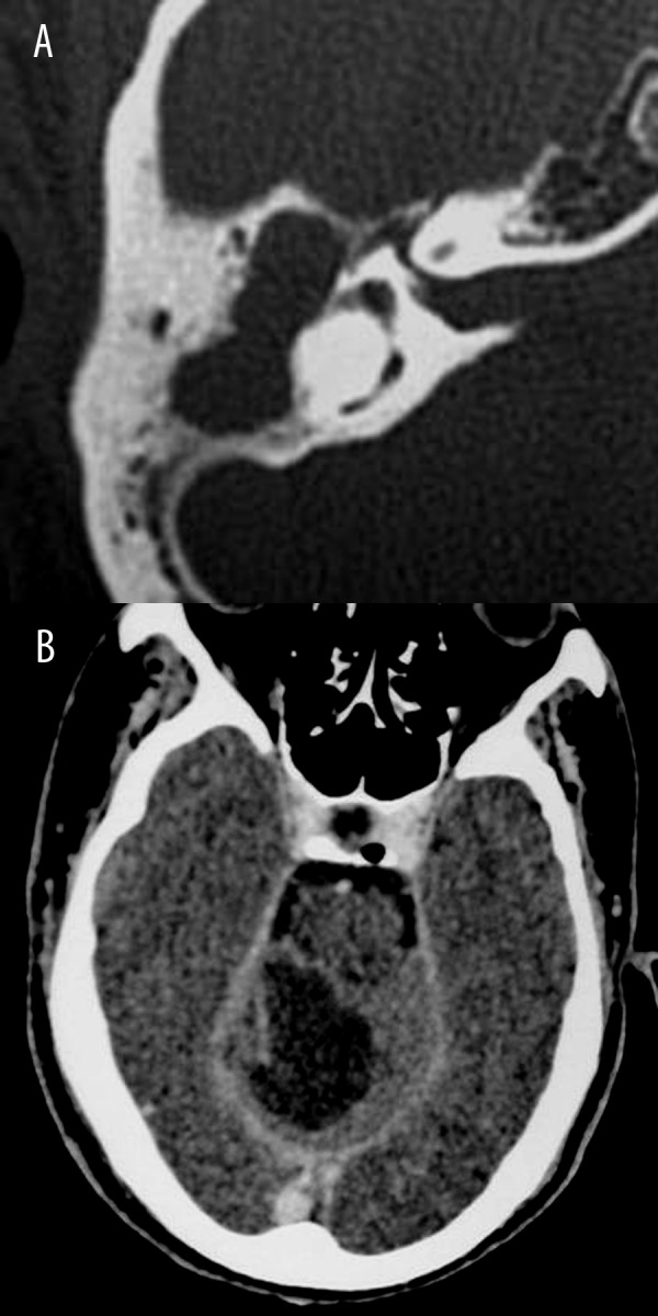
Material/methods: Prospective study was conducted on 61 consecutive patients (mean age 26.8 years) with middle ear cholesteatoma. CT scan of the temporal bone and surgery were performed in all patients. CT staging classified cholesteatoma according to its location in the tympanic cavity (T); extension into the mastoid (M); and associated complications (C). Cholesteatoma was staged as stage I (T1, T2), stage II (T3, M1, M2, C1), and stage III (C2).
Results: The overall sensitivity of CT staging of cholesteatoma compared to surgery was 88% with excellent agreement and correlation between CT findings and intra-operative findings (K=0.863, r=0.86, P=0.001). There was excellent agreement and correlation of CT staging with surgical findings for T location (K=0.811, r=0.89, P=0.001), good for M extension (K=0.734, r=0.88, P=0.001), and excellent for associated C complications (K=1.00, r=1.0, P=0.001). Atticotympanotomy was carried out in stage I (n=14), intact canal wall surgery was performed in stage II (n=38), and canal wall down surgery was done in stage III (n=5) and stage II (n=4).
Conclusions: We established CT staging of middle ear cholesteatoma that helps surgeons to select an appropriate surgery.
Keywords: Adenovirus Infections, Human; Anatomy, Comparative; Imaging, Three-Dimensional; Pain.
Figures
References
-
- Juliano AF, Ginat DT, Moonis G. Imaging review of the temporal bone: part I. Anatomy and inflammatory and neoplastic processes. Radiology. 2013;269:17–33. - PubMed
-
- Más-Estellés F, Mateos-Fernández M, Carrascosa-Bisquert B, et al. Contemporary non-echo-planar diffusion-weighted imaging of middle ear cholesteatomas. Radiographics. 2012;32:1197–213. - PubMed
-
- Lemmerling M, De Foer B, Verbist B, VandeVyver V. Imaging of inflammatory and infectious diseases in the temporal bone. Neuroimag Clin North Am. 2009;19:321–37. - PubMed
-
- Bruce B, Ian G. Acquired cholesteatoma: classification and outcomes. Otol Neurotol. 2011;32:992–95. - PubMed
LinkOut - more resources
Full Text Sources
Miscellaneous