. 2015 May 20:6:1183-91.
doi: 10.3762/bjnano.6.120. eCollection 2015.

Tattoo ink nanoparticles in skin tissue and fibroblasts

Affiliations

Tattoo ink nanoparticles in skin tissue and fibroblasts

Colin A Grant et al. Beilstein J Nanotechnol. .

Abstract

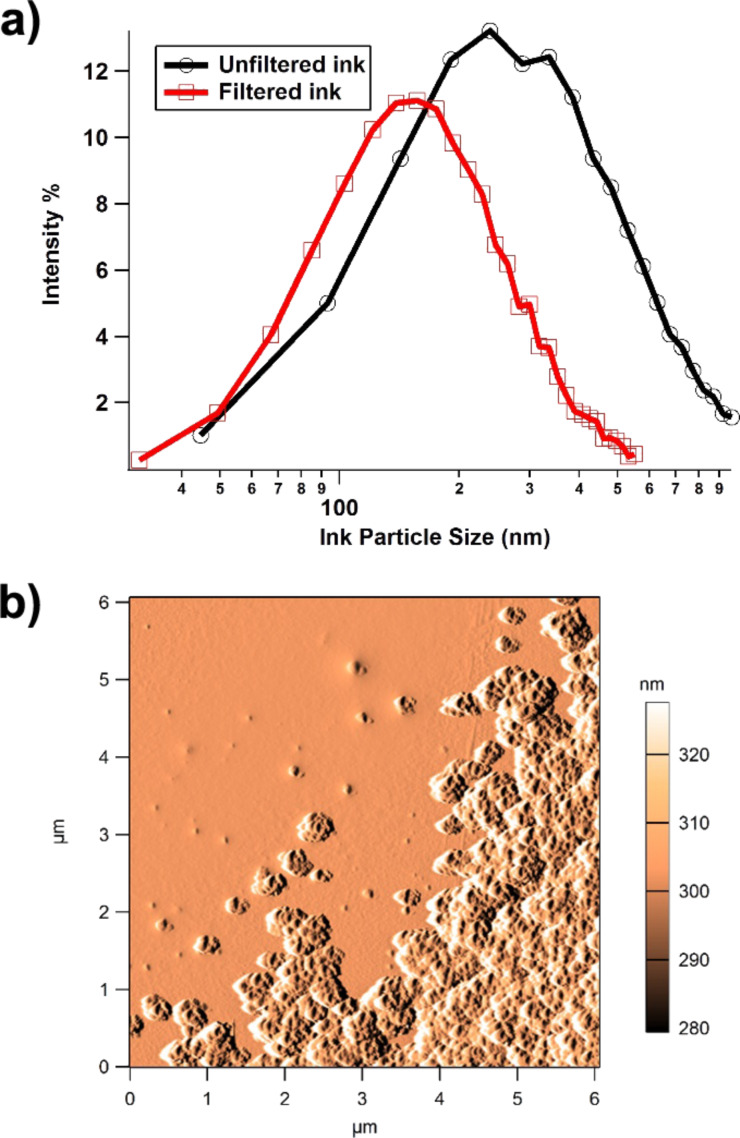
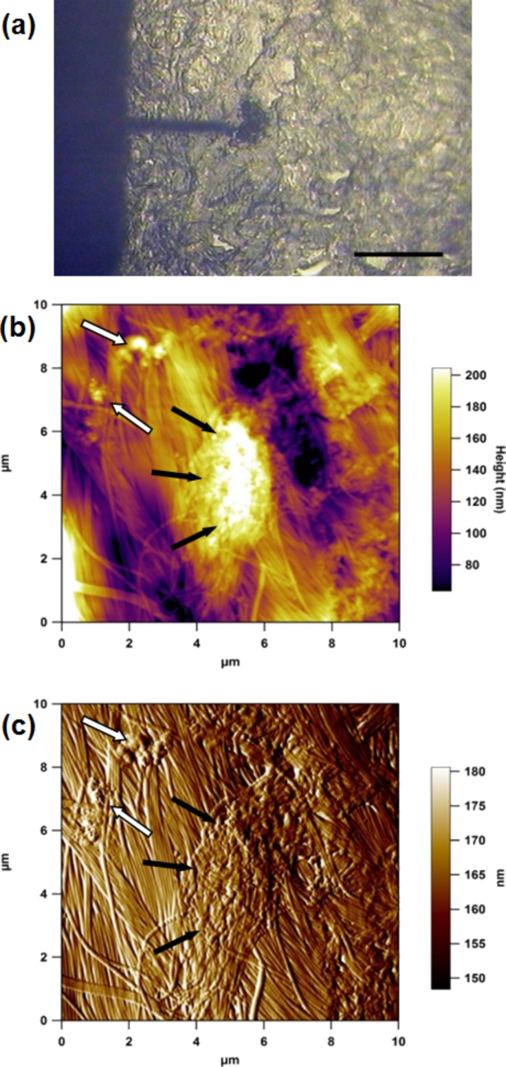
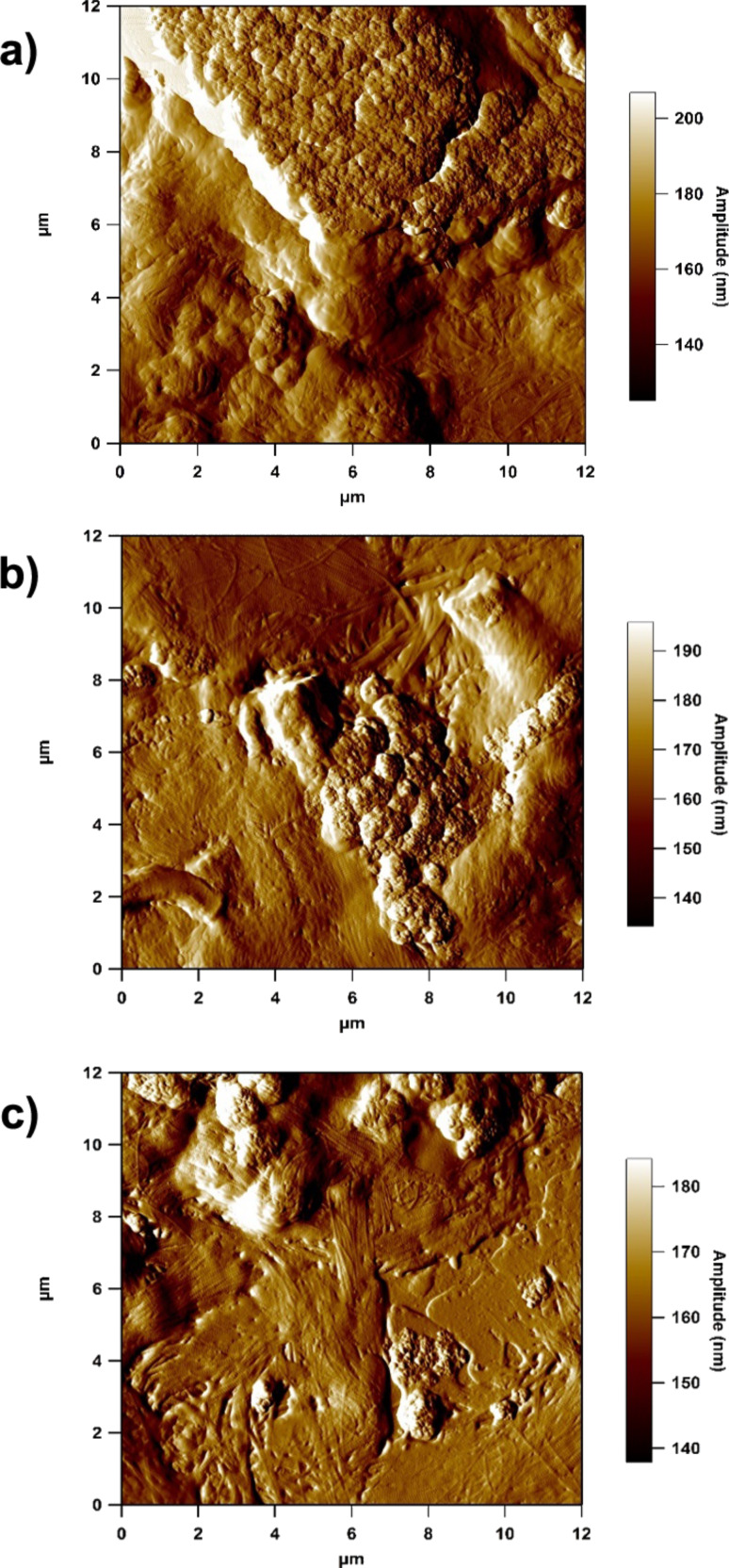
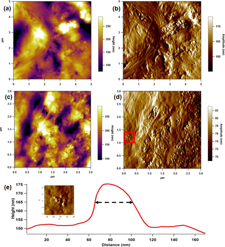
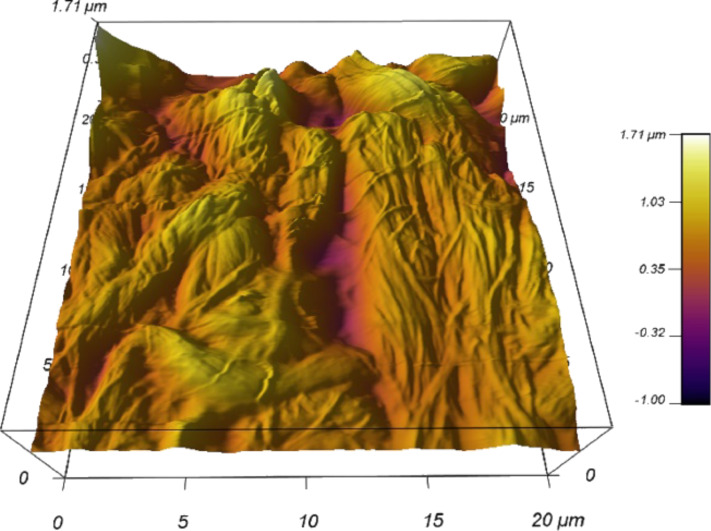
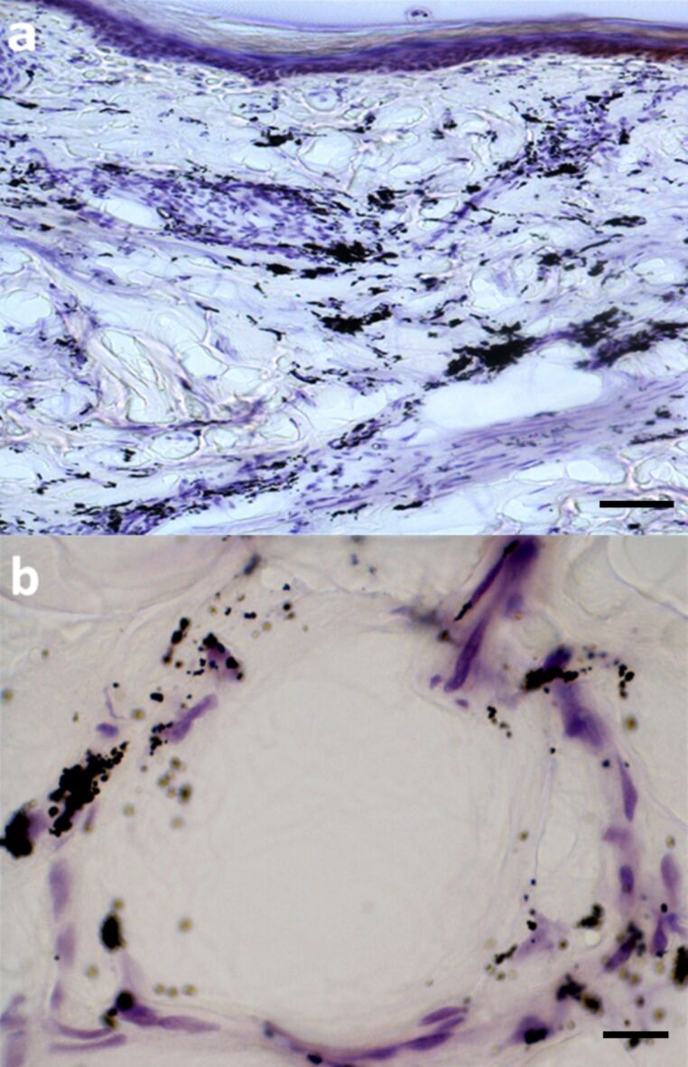
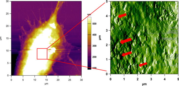
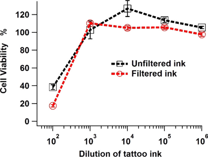
Tattooing has long been practised in various societies all around the world and is becoming increasingly common and widespread in the West. Tattoo ink suspensions unquestionably contain pigments composed of nanoparticles, i.e., particles of sub-100 nm dimensions. It is widely acknowledged that nanoparticles have higher levels of chemical activity than their larger particle equivalents. However, assessment of the toxicity of tattoo inks has been the subject of little research and ink manufacturers are not obliged to disclose the exact composition of their products. This study examines tattoo ink particles in two fundamental skin components at the nanometre level. We use atomic force microscopy and light microscopy to examine cryosections of tattooed skin, exploring the collagen fibril networks in the dermis that contain ink nanoparticles. Further, we culture fibroblasts in diluted tattoo ink to explore both the immediate impact of ink pigment on cell viability and also to observe the interaction between particles and the cells.

Keywords: atomic force microscopy (AFM); dermis; nanoparticles; skin; tattoo ink.


Figures

Figure 1
Figure 1
(a) Particle size distribution of filtered vs unfiltered commercially available tattoo ink, showing data ranging between 30 to 600 nm (filtered) and 40 to 970 nm (unfiltered). (b) Amplitude image of tattoo ink particles showing single and agglomerated particles adhered to a glass substrate.
Figure 2
Figure 2
(a) AFM optical image (10×) showing the cantilever over a region of tattoo ink in the dermis; scale bar 200 μm. (b) 10 μm AFM height and (c) amplitude (error) image of cryosectioned tattooed skin. Black arrows indicate a large agglomerate and white arrows smaller agglomerates of tattoo ink particles.
Figure 3
Figure 3
12 μm amplitude images of highly agglomerated tattoo ink particles in the collagen network.
Figure 4
Figure 4
Height (a & c) and amplitude (b & d) images of disperse ink particles in the dermal collagen network. (Inset of (e)) 500 nm image of small cluster of ink particles from the solid red square (e) Line profile showing a particle of 37.5 nm width at half height.
Figure 5
Figure 5
3D reconstruction of AFM image on non-tattoo portion of the skin tissue under aqueous conditions (UHQ water).
Figure 6
Figure 6
Light microscopy view of stained adult human tattooed arm skin. (a) Large deposits of dark ink particles distributed in a clumped manner in the dermis; scale bar 75 μm. (b) A deep dermal vessel with aggregations of ink particles in/around vessel wall and inside some associated cells; scale bar 15 μm.
Figure 7
Figure 7
(a) AFM height image of a large fibroblast in vitro and incubated with diluted tattoo ink (1:10,000), followed by chemical fixation. (b) Phase image (5 μm) of the highest part of the cell body, which shows many regions (red arrows) of clumped ink particles on the cell surface.
Figure 8
Figure 8
Cell viability of skin dermal fibroblasts incubated in diluted tattoo ink for seven days (filtered and unfiltered).

References

    1. Pabst M A, Letofsky-Papst I, Bock E, Moser M, Dorfer L, Egarter-Vigl E, Hofer F. J Archaeol Sci. 2009;36:2335–2341. doi: 10.1016/j.jas.2009.06.016. - DOI
    1. Dorfer L, Moser M, Bahr F, Spindler K, Egarter-Vigl E, Giullén S, Dohr G, Kenner T. Lancet. 1999;354:1023–1025. doi: 10.1016/S0140-6736(98)12242-0. - DOI - PubMed
    1. Zirkin H J, Avinoach I, Edelwitz P. Cutis. 2001;67:471–472. - PubMed
    1. Regensburger J, Lehner K, Maisch T, Vasold R, Santarelli F, Engel E, Gollmer A, König B, Landthaler M, Bäumler W. Exp Dermatol. 2010;19:e275–e281. doi: 10.1111/j.1600-0625.2010.01068.x. - DOI - PubMed
    1. Høgsberg T, Hutton Carlsen K, Serup J. J Eur Acad Dermatol Venereol. 2013;27:846–852. doi: 10.1111/j.1468-3083.2012.04590.x. - DOI - PubMed

LinkOut - more resources