

Cytomegalovirus Infection Triggers the Secretion of the PPARγ Agonists 15-Hydroxyeicosatetraenoic Acid (15-HETE) and 13-Hydroxyoctadecadienoic Acid (13-HODE) in Human Cytotrophoblasts and Placental Cultures
- PMID: 26171612
- PMCID: PMC4501751
- DOI: 10.1371/journal.pone.0132627
Cytomegalovirus Infection Triggers the Secretion of the PPARγ Agonists 15-Hydroxyeicosatetraenoic Acid (15-HETE) and 13-Hydroxyoctadecadienoic Acid (13-HODE) in Human Cytotrophoblasts and Placental Cultures
Abstract
Introduction: Congenital infection by human cytomegalovirus (HCMV) is a leading cause of congenital abnormalities of the central nervous system. Placenta infection by HCMV allows for viral spread to fetus and may result in intrauterine growth restriction, preeclampsia-like symptoms, or miscarriages. We previously reported that HCMV activates peroxisome proliferator-activated receptor gamma (PPARγ) for its own replication in cytotrophoblasts. Here, we investigated the molecular bases of PPARγ activation in infected cytotrophoblasts.
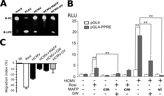
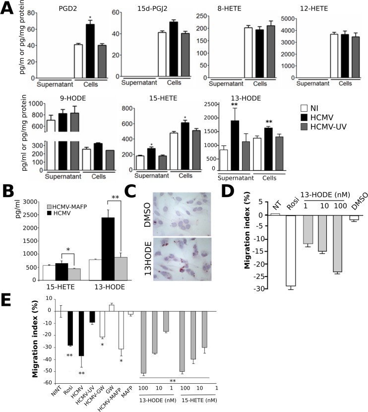
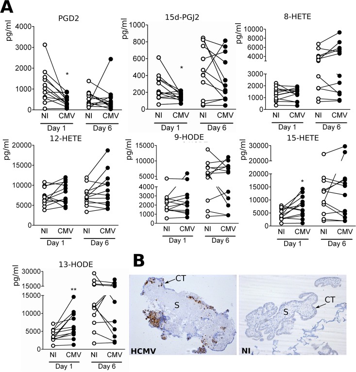
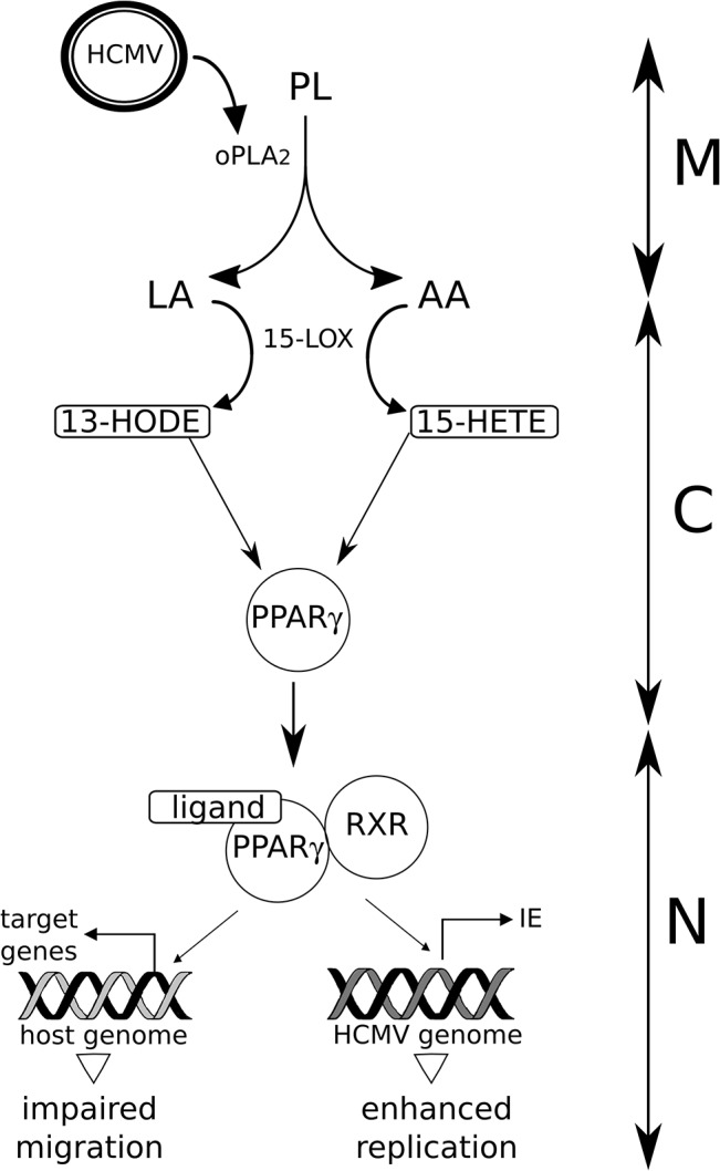
Results: We show that onboarded cPLA2 carried by HCMV particles is required for effective PPARγ activation in infected HIPEC cytotrophoblasts, and for the resulting inhibition of cell migration. Natural PPARγ agonists are generated by PLA2 driven oxidization of linoleic and arachidonic acids. Therefore, using HPLC coupled with mass spectrometry, we disclosed that cellular and secreted levels of 13-hydroxyoctadecadienoic acid (13-HODE) and 15-hydroxyeicosatetraenoic acid (15-HETE) were significantly increased in and from HIPEC cytotrophoblasts at soon as 6 hours post infection. 13-HODE treatment of uninfected HIPEC recapitulated the effect of infection (PPARγ activation, migration impairment). We found that infection of histocultures of normal, first-term, human placental explants resulted in significantly increased levels of secreted 15-HETE and 13-HODE.
Conclusion: Our findings reveal that 15-HETE and 13-HODE could be new pathogenic effectors of HCMV congenital infection They provide a new insight about the pathogenesis of congenital infection by HCMV.
Conflict of interest statement
Figures
Similar articles
-
PPARγ Is Activated during Congenital Cytomegalovirus Infection and Inhibits Neuronogenesis from Human Neural Stem Cells.PLoS Pathog. 2016 Apr 14;12(4):e1005547. doi: 10.1371/journal.ppat.1005547. eCollection 2016 Apr. PLoS Pathog. 2016. PMID: 27078877 Free PMC article.
-
15-Lipoxygenases and its metabolites 15(S)-HETE and 13(S)-HODE in the development of non-small cell lung cancer.Thorax. 2010 Apr;65(4):321-6. doi: 10.1136/thx.2009.122747. Thorax. 2010. PMID: 20388757
-
Antineoplastic effects of 15(S)-hydroxyeicosatetraenoic acid and 13-S-hydroxyoctadecadienoic acid in non-small cell lung cancer.Cancer. 2015 Sep 1;121 Suppl 17:3130-45. doi: 10.1002/cncr.29547. Cancer. 2015. PMID: 26331820
-
Congenital cytomegalovirus infection undermines early development and functions of the human placenta.Placenta. 2017 Nov;59 Suppl 1:S8-S16. doi: 10.1016/j.placenta.2017.04.020. Epub 2017 Apr 25. Placenta. 2017. PMID: 28477968 Review.
-
The Role of Arachidonic and Linoleic Acid Derivatives in Pathological Pregnancies and the Human Reproduction Process.Int J Mol Sci. 2020 Dec 17;21(24):9628. doi: 10.3390/ijms21249628. Int J Mol Sci. 2020. PMID: 33348841 Free PMC article. Review.
Cited by
-
PPARγ Is Activated during Congenital Cytomegalovirus Infection and Inhibits Neuronogenesis from Human Neural Stem Cells.PLoS Pathog. 2016 Apr 14;12(4):e1005547. doi: 10.1371/journal.ppat.1005547. eCollection 2016 Apr. PLoS Pathog. 2016. PMID: 27078877 Free PMC article.
-
The Role of Congenital Cytomegalovirus Infection in Adverse Birth Outcomes: A Review of the Potential Mechanisms.Viruses. 2020 Dec 24;13(1):20. doi: 10.3390/v13010020. Viruses. 2020. PMID: 33374185 Free PMC article. Review.
-
Immune mechanisms affected by cyclooxygenase inhibition combined with antiviral treatment in calves infected with bovine respiratory syncytial virus.PLoS One. 2025 Apr 22;20(4):e0321642. doi: 10.1371/journal.pone.0321642. eCollection 2025. PLoS One. 2025. PMID: 40261882 Free PMC article.
-
Human Cytomegalovirus Modifies Placental Small Extracellular Vesicle Composition to Enhance Infection of Fetal Neural Cells In Vitro.Viruses. 2022 Sep 13;14(9):2030. doi: 10.3390/v14092030. Viruses. 2022. PMID: 36146834 Free PMC article.
-
Machine learning and semi-targeted lipidomics identify distinct serum lipid signatures in hospitalized COVID-19-positive and COVID-19-negative patients.Metabolism. 2022 Jun;131:155197. doi: 10.1016/j.metabol.2022.155197. Epub 2022 Apr 2. Metabolism. 2022. PMID: 35381232 Free PMC article.
References
Publication types
MeSH terms
Substances
LinkOut - more resources
Full Text Sources
Other Literature Sources
