. 2015 Aug 11;113(4):653-9.
doi: 10.1038/bjc.2015.251. Epub 2015 Jul 14.

Impact of p16 status on pro- and anti-angiogenesis factors in head and neck cancers

Affiliations

Impact of p16 status on pro- and anti-angiogenesis factors in head and neck cancers

P Baruah et al. Br J Cancer. .

Abstract

Background: Head and neck cancers (HNC) are aggressive tumours. Overexpression of p16 in HNC correlates with human papilloma virus (HPV)-associated HNC that carry a better prognosis than HPV-negative tumours. Angiogenesis is an important factor in tumour progression. Our aim was to dissect the impact of p16 expression on angiogenesis factors in HNC.

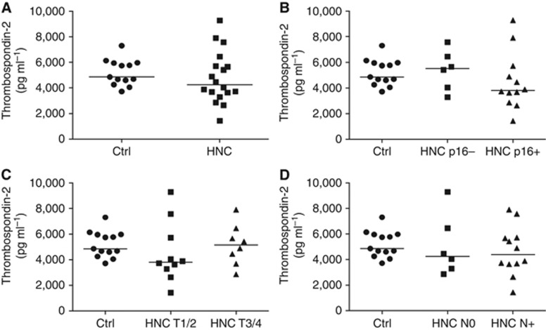
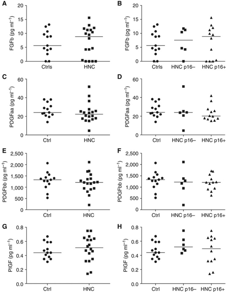
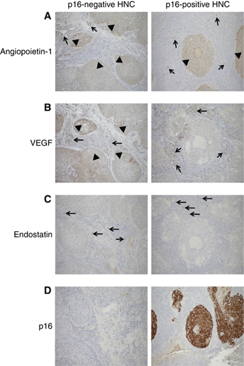
Methods: Eighteen newly diagnosed HNC patients and controls were analysed. Eleven pro- and anti-angiogenesis factors were quantified using multiplex ELISA in HNC patients and controls. Angiogenesis factors were analysed in tumour tissue using immunohistochemistry.

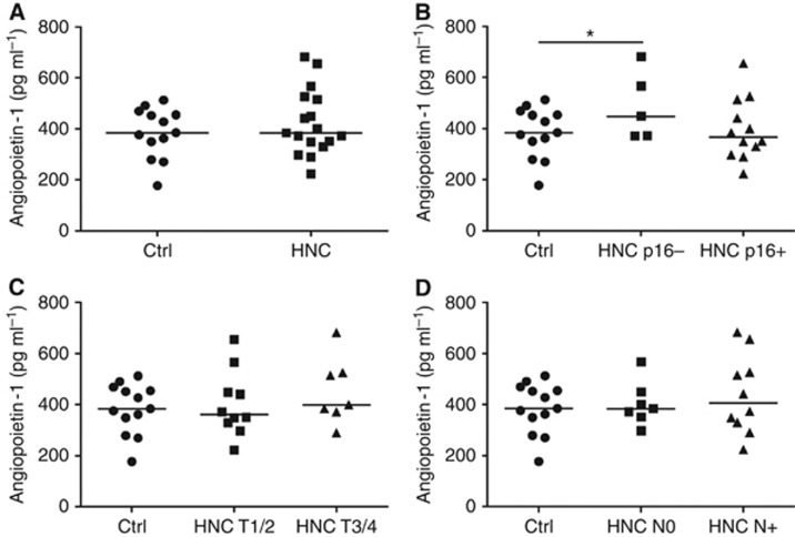
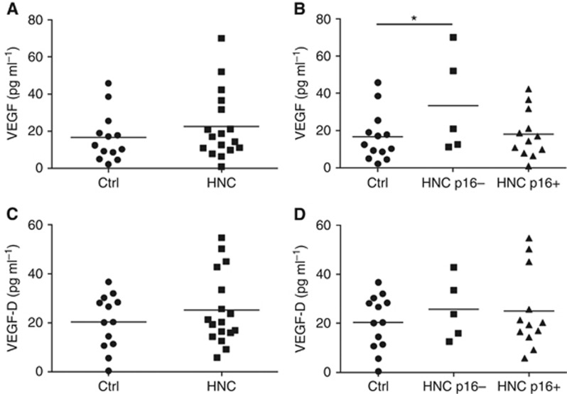
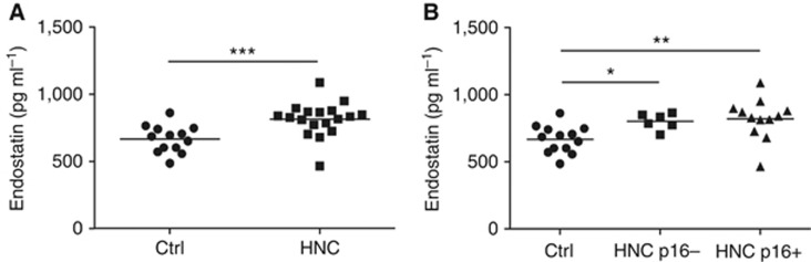
Results: Circulating levels of endostatin (anti-angiogenesis factor) were higher in the HNC group compared with healthy donors. Interestingly, the pro-angiogenesis factors angiopoietin-1 and vascular endothelial growth factor (VEGF) were significantly higher in patients with p16-negative compared with p16-positive HNC. Moreover, the major source of VEGF in p16-positive HNC tissue was tumour stromal cells. In contrast, both tumour cells and stromal cells expressed VEGF in p16-negative tissue.

Conclusions: We show that p16-negative tumours associate with increased circulating levels of pro-angiogenic VEGF and angiopoietin-1. Tissue expression of VEGF differs between p16-positive and p16-negative tumours. These findings may explain differences in the biological behaviour of p16-positive and p16-negative HNC. Better understanding of mechanisms by which the p16 status influences tumour angiogenesis may guide the development of targeted therapies.


Figures

Figure 1
Figure 1
Angiopoietin-1 levels in patients with HNC and controls. Circulating levels of angiopoietin-1 were quantified using multiplex ELISA in sera from healthy individuals (Ctrl, n=13) and patients with HNC (n=17). (A) The scatter plots show angiopoietin-1 concentration in the two study groups (horizontal bars show the mean value for each group). (B) Angiopoietin-1 levels were compared between p16-positive (HNC p16+) or p16-negative (HNC p16−) HNC patients and controls. *P<0.05. (C) The plots display angiopoietin-1 in patients with early tumour stage (T1/2), advanced tumour stage (T3/4) and controls. (D) The scatter plots display angiopoietin-1 in patients without nodal disease (N0), nodal disease (N+) and controls.
Figure 2
Figure 2
VEGF levels in patients with HNC and controls. Circulating levels of VEGF were quantified using multiplex ELISA in sera from healthy individuals (Ctrl, n=13) and patients with HNC (n=17). (A) The scatter plots show VEGF concentration in the two study groups (horizontal bars show the mean value for each group). (B) VEGF levels were compared between p16-positive (HNC p16+) or p16-negative (HNC p16−) HNC patients and controls. *P<0.05. (C) The plots display VEGF-D in the two study groups. (D) The scatter plots display VEGF-D in p16-positive (HNC p16+) and negative (HNC p16−) patients and controls. VEGF=vascular endothelial growth factor.
Figure 3
Figure 3
Endostatin levels in patients with HNC and controls. Circulating levels of endostatin were quantified using multiplex ELISA in sera from healthy individuals (Ctrl, n=13) and patients with HNC (n=18). (A) The scatter plots show endostatin concentration in the two study groups (horizontal bars show the mean value for each group). (B) Endostatin levels were compared between p16-positive (HNC p16+) or p16-negative (HNC p16−) HNC patients and controls. ***P<0.001, **P<0.01, *P<0.05.
Figure 4
Figure 4
Thrombospondin levels in patients with HNC and controls. Circulating levels of thrombospondin-2 were quantified using multiplex ELISA in sera from healthy individuals (Ctrl, n=13) and patients with HNC (n=18). (A) The scatter plots show thrombospondin-2 concentration in the two study groups (horizontal bars show the mean value for each group). (B) Thrombospondin-2 levels were compared between p16-positive (HNC p16+) or p16-negative (HNC p16−) HNC patients and controls. (C) The plots display thrombospondin-2 in patients with early tumour stage (T1/2), advanced tumour stage (T3/4) and controls. (D) The scatter plots display thrombospondin-2 in patients without nodal disease (N0), nodal disease (N+) and controls.
Figure 5
Figure 5
Other angiogenic factor levels in patients with HNC and controls. Circulating levels of angiogenic factors were quantified using multiplex ELISA in sera from healthy individuals (Ctrl, n=13) and patients with HNC (n=18). (A) The scatter plots show FGF-b concentration in controls and HNC patients. Horizontal bars show the mean value for each group. (B) FGF-b levels in p16-positive (HNC p16+) or p16-negative (HNC p16−) HNC patients and controls. (C) The scatter plots show PDGF-aa concentration in controls and HNC patients. (D) PDGF-aa levels in p16-positive or p16-negative HNC patients and controls. (E) The scatter plots show PDGF-bb concentration of controls and HNC patients. (F) PDGF-bb levels in p16-positive or p16-negative HNC patients and controls. (G) The scatter plots show PlGF concentration controls and HNC patients. (H) PlGF levels in p16-positive or p16-negative HNC patients and controls. FGF-b=fibroblast growth factor-basic; PDGF=platelet-derived growth factor; PlGF=placental growth factor.
Figure 6
Figure 6
Expression of angiopoietin-1, VEGF and endostatin in p16-negative and p16-positive HNC tissue. Expression of angiogenic factors (angiopoietin-1, VEGF and endostatin) was assessed in tissue from five patients with p16-negative and five patients with p16-positive HNC using immunohistochemistry (see Materials and Methods for details). (A) Angiopoietin-1 expression in tumour cells (arrow heads) and tumour stroma (arrows). (B) VEGF expression in tumour tissue (arrow heads) and stromal tissue (arrows). (C) Expression of endostatin in the tumour stroma (arrows). (D) p16 staining in the tumour tissue used for analysis of angiogenesis factor expression.

References

    1. Ang KK, Harris J, Wheeler R, Weber R, Rosenthal DI, Nguyen-Tan PF, Westra WH, Chung CH, Jordan RC, Lu C, Kim H, Axelrod R, Silverman CC, Redmond KP, Gillison ML (2010) Human papillomavirus and survival of patients with oropharyngeal cancer. N Engl J Med 363(1): 24–35. - PMC - PubMed
    1. Blezinger P, Wang J, Gondo M, Quezada A, Mehrens D, French M, Singhal A, Sullivan S, Rolland A, Ralston R, Min W (1999) Systemic inhibition of tumor growth and tumor metastases by intramuscular administration of the endostatin gene. Nat Biotechnol 17(4): 343–348. - PubMed
    1. Cohen EE, Davis DW, Karrison TG, Seiwert TY, Wong SJ, Nattam S, Kozloff MF, Clark JI, Yan DH, Liu W, Pierce C, Dancey JE, Stenson K, Blair E, Dekker A, Vokes EE (2009) Erlotinib and bevacizumab in patients with recurrent or metastatic squamous-cell carcinoma of the head and neck: a phase I/II study. Lancet Oncol 10(3): 247–257. - PMC - PubMed
    1. Ferrara N, Davis-Smyth T (1997) The biology of vascular endothelial growth factor. Endocr Rev 18(1): 4–25. - PubMed
    1. Folkman J, Watson K, Ingber D, Hanahan D (1989) Induction of angiogenesis during the transition from hyperplasia to neoplasia. Nature 339(6219): 58–61. - PubMed

Publication types

MeSH terms