. 2015 Sep:271:445-56.
doi: 10.1016/j.expneurol.2015.07.001. Epub 2015 Jul 11.

JAK/STAT pathway regulation of GABAA receptor expression after differing severities of experimental TBI

Affiliations

JAK/STAT pathway regulation of GABAA receptor expression after differing severities of experimental TBI

Daniel J Raible et al. Exp Neurol. 2015 Sep.

Abstract

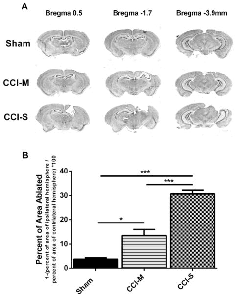
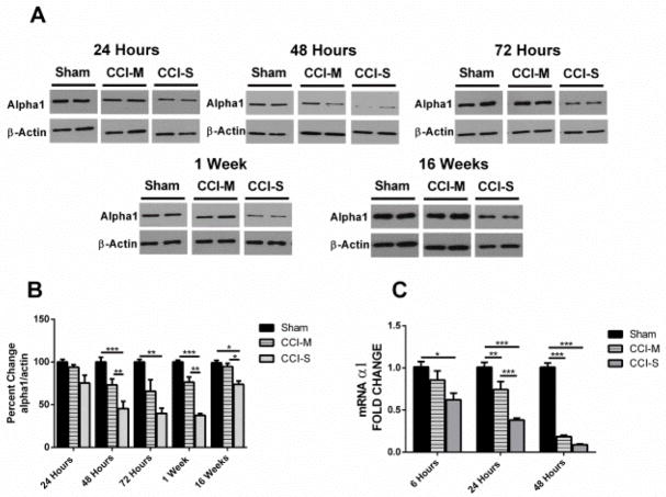
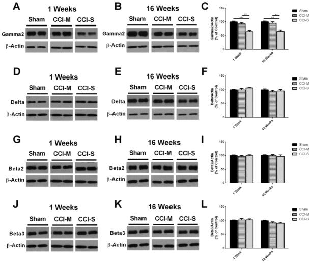
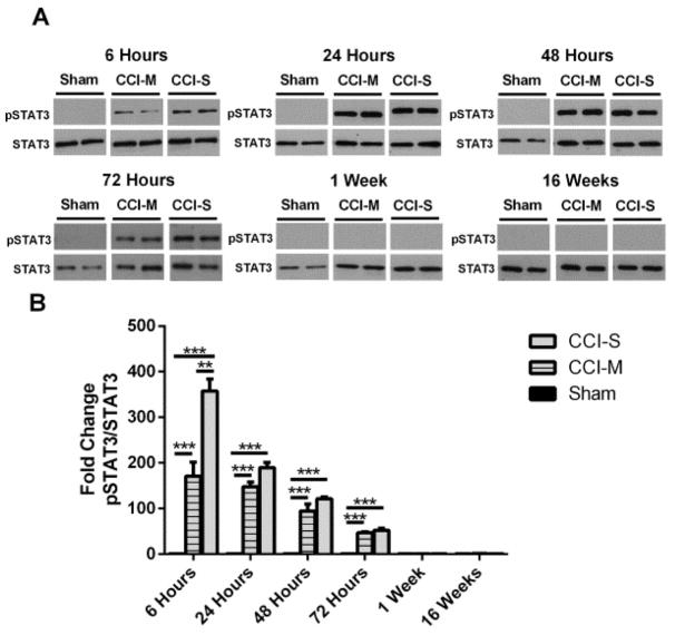
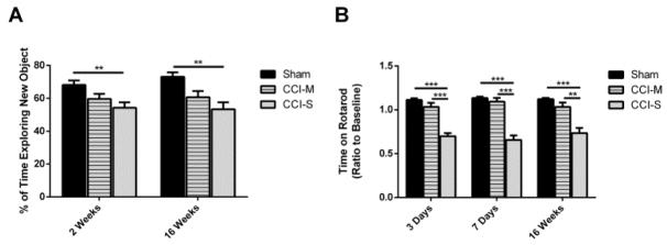
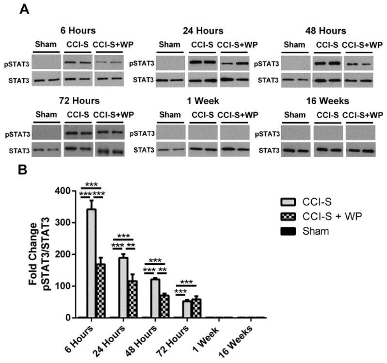
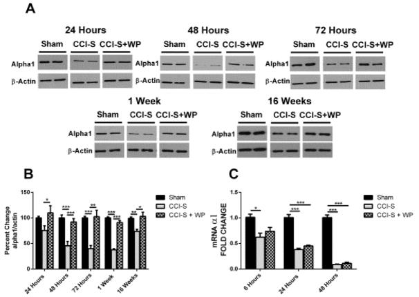
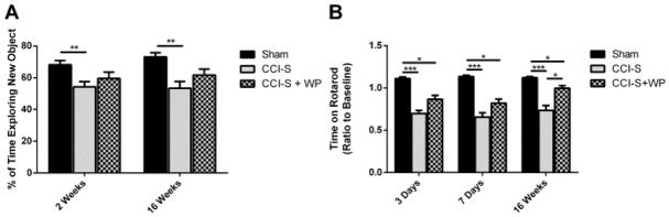
Synaptic inhibition in the adult brain is primarily mediated by the γ-aminobutyric acid (GABA) type A receptor (GABA(A)R). The distribution, properties, and dynamics of these receptors are largely determined by their subunit composition. Alteration of subunit composition after a traumatic brain injury (TBI) may result in abnormal increased synaptic firing and possibly contribute to injury-related pathology. Several studies have shown that the Janus Kinase/Signal Transducer and Activator of Transcription (JAK/STAT) signaling pathway can alter GABA(A)R subunit expression. The present study investigated changes in JAK/STAT pathway activation after two different severities of experimental TBI in the mouse using the controlled cortical impact (CCI) model. It also investigated whether modulating the activation of the JAK/STAT pathway after severe controlled cortical impact (CCI-S) with a JAK/STAT inhibitor (WP1066) alters post-traumatic epilepsy development and/or neurological recovery after injury. Our results demonstrated differential changes in both the activation of STAT3 and the expression of the GABA(A)R α1 and γ2 subunit levels that were dependent on the severity of the injury. The change in the GABA(A)R α1 subunit levels appeared to be at least partly transcriptionally mediated. We were able to selectively reverse the decrease in GABA(A)R α1 protein levels with WP1066 treatment after CCI injury. WP1066 treatment also improved the degree of recovery of vestibular motor function after injury. These findings suggest that the magnitude of JAK/STAT pathway activation and GABA(A)R α1 subunit level decrease is dependent on injury severity in this mouse model of TBI. In addition, reducing JAK/STAT pathway activation after severe experimental TBI reverses the decrease in the GABA(A)R α1 protein levels and improves vestibular motor recovery.

Keywords: GABA(A) receptor; JAK/STAT pathway; STAT3; Traumatic brain injury.


Figures

Figure 1
Figure 1
Progressive tissue loss following CCI-S and CCI-M injuries. (A) A series of representative images of CV stained coronal sections from sham, CCI-S and CCI-M injured mice. Sections show cerebral damage at three different levels posterior from bregma. Calibration bar, 5 mm (B) Quantitation of cerebral extent by measuring the area of the ipsilateral hemisphere and normalizing it to the area of the contralateral hemisphere for sham, CCI-S and CCI-M injured mice. Each group is statistically different from each other group with CCI-S having the largest lesion. * P < 0.05, ** P < 0.01, P < 0.001 (n = 4 for sham, n = 7 for CCI-M and n = 8 for CCI-S)
Figure 2
Figure 2
Temporal profile of GABAA α1 receptor subunit after CCI. (A) Representative western blots form whole hippocampus of mice 24 hours, 48 hours, 72 hours, 1 week and 16 weeks after CCI probed with anti-GABAAR α1 and β-actin antibodies. (B) Quantification of α1 blots showed a significant decrease at 48 hours through 16 weeks after CCI-S injury relative to CCI-M and sham injured controls. α1 levels were normalized to β-actin levels and expressed as a percent change compared to sham injured controls. (C) mRNA levels of α1 were quantified using RT-PCR analysis and represented as histograms showing the fold change of α1 6 hours, 24 hours and 48 hours after CCI in CCI-S, CCI-M and sham injured controls. α1 mRNA levels were normalized to B2M mRNA levels in the same samples and expressed as a fold change compared to sham injured controls (defined as 1). * P < 0.05, ** P < 0.01, P < 0.001 (for A and B n = 7 for sham, n = 7 for CCI-S and n = 6 for CCI-M at 24 hours and n = 7 for sham, n = 7 for CCI-S and n = 7 for CCI-M at 48 hours and n = 6 for sham, n = 6 for CCI-S and n = 6 for CCI-M at 72 hours and n = 6 for sham, n = 7 for CCI-S and n = 7 for CCI-M at 1 week and n = 7 for sham, n = 10 for CCI-S and n = 9 for CCI-M at 16 weeks) (for C n = 7 for sham, n = 7 for CCI-S and n = 6 for CCI-M at 6 hours and n = 7 for sham, n = 7 for CCI-S and n = 7 for CCI-M at 24 hours and n = 7 for sham, n = 7 for CCI-S and n = 7 for CCI-M at 48 hours).
Figure 3
Figure 3
GABAA receptor subunit γ2 is decreased while δ, β2 and β3 are unchanged 1 and 16 weeks post CCI. (A) Representative western blots from whole hippocampus of mice 1 week after CCI probed with anti-GABAAR γ2 and β-actin antibodies. (B) Representative western blots from whole hippocampus of mice 16 weeks after CCI probed with anti-GABAAR γ2 and β-actin antibodies. (C) Quantification of γ2 blots showed that the γ2 subunit is significantly decreased 1 and 16 weeks after CCI-S relative to CCI-M and sham injured controls. (D) Representative western blots from whole hippocampus of mice 1 week after CCI probed with anti-GABAAR δ and β-actin antibodies. (E) Representative western blots from whole hippocampus of mice 16 weeks after CCI probed with anti-GABAAR δ and β-actin antibodies. (F) Quantification of δ blots showed that the δ subunit was unchanged 1 and 16 weeks after CCI-S relative to CCI-M and sham injured controls. (G) Representative western blots from whole hippocampus of mice 1 week after CCI probed with anti-GABAAR β2 and β-actin antibodies. (H) Representative western blots from whole hippocampus of mice 16 weeks after CCI probed with anti-GABAAR β2 and β-actin antibodies. (I) Quantification of β2 blots shows that the β2 subunit was unchanged 1 and 16 weeks after CCI-S relative to CCI-M and sham injured controls. (J) Representative western blots from whole hippocampus of mice 1 week after CCI probed with anti-GABAAR β3 and β-actin antibodies. (K) Representative western blots from whole hippocampus of mice 16 weeks after CCI probed with anti-GABAAR β3 and β-actin antibodies. (L) Quantification of β3 blots showed that the β3 subunit was unchanged 1 and 16 weeks after CCI-S relative to CCI-M and sham injured controls. * P < 0.05, ** P < 0.01, P < 0.001 (n = 6 for sham, n = 7 for CCI-S and n = 7 for CCI-M at 1 week and n = 10 for sham, n = 10 for CCI-S and n = 9 for CCI-M at 16 weeks).
Figure 4
Figure 4
Phosphorylated STAT3 levels in injured hippocampus after CCI. (A) Representative western blots of protein homogenates from whole ipsilateral hippocampus (relative to side of CCI) of mice 6 hours, 24 hours, 48 hours, 1 week and 16 weeks after CCI probed with pSTAT3 and STAT3 antibodies. (B) Quantification of pSTAT3 levels from CCI-S, CCI-M and sham injured controls showed that from 6 hours to 72 hours post injury the pSTAT3 levels were higher in both the CCI-S and CCI-M injured group when compared to sham injured controls. Levels of pSTAT3 in the CCI-S groups were statistically higher than the levels in the CCI-M group at 6 hours post injury but, at all other time points there was no statistical difference between CCI-S and CCI-M groups in pSTAT3 protein levels. * P < 0.05, ** P < 0.01, P < 0.001 (n = 9 for sham, n = 9 for CCI-S and n = 9 for CCI-M at 6 hours, n = 10 for sham, n = 9 for CCI-S and n = 10 for CCI-M at 24 hours and n = 7 for sham, n = 7 for CCI-S and n = 7 for CCI-M at 48 hours and n = 6 for sham, n = 8 for CCI-S and n = 6 for CCI-M at 72 hours and n = 6 for sham, n = 7 for CCI-S and n = 7 for CCI-M at 1 week and n = 6 for sham, n = 7 for CCI-S and n = 6 for CCI-M at 16 weeks).
Figure 5
Figure 5
Memory and vestibular motor performance is significantly reduced after CCI-S. (A) Quantification of average time spent exploring the new object during Novel Object Recognition testing shows that the CCI-S injured mice performed statistically worse than the sham injured controls or CCI-M injured mice. (B) Quantification of average time spent on the Rotarod apparatus showed that the CCI-S injured mice performed significantly worse than the CCI-M injured mice or sham injured controls. * P < 0.05, ** P < 0.01, P < 0.001 (for A n = 19 for sham, n = 18 for CCI-S and n = 12 for CCI-M at 2 weeks and n = 11 for sham, n = 12 for CCI-S and n = 10 for CCI-M at 16 weeks) (for B n = 20 for sham, n = 28 for CCI-S and n = 19 for CCI-M at 3 days and n = 20 for sham, n = 28 for CCI-S and n = 19 for CCI-M at 7 days and n = 12 for sham, n = 13 for CCI-S and n = 9 for CCI-M at 16 weeks).
Figure 6
Figure 6
WP1066 administration after CCI-S reduces phosphorylated STAT3 levels in injured hippocampus. (A) Representative western blots of protein homogenates from whole ipsilateral hippocampus (relative to side of CCI) of mice 6 hours, 24 hours, 48 hours, 1 week and 16 weeks after CCI-S treated with vehicle or WP1066 probed with pSTAT3 and STAT3 antibodies. (B) Quantification of pSTAT3 levels from CCI-S, CCI-S + WP and sham injured controls showed that from 6 hours to 48 hours post injury that pSTAT3 levels were statistically lower than CCI-S injured mice treated with WP1066 compared to vehicle-treated CCI-S mice. * P < 0.05, ** P < 0.01, P < 0.001 (n = 9 for sham, n = 9 for CCI-S and n = 6 for CCI-S + WP at 6 hours, n = 10 for sham, n = 9 for CCI-S and n = 7 for CCI-S + WP at 24 hours and n = 7 for sham, n = 7 for CCI-S and n = 7 for CCI-S + WP at 48 hours and n = 6 for sham, n = 8 for CCI-S and n = 6 for CCI-S + WP at 72 hours and n = 6 for sham, n = 7 for CCI-S and n = 6 for CCI-S + WP at 1 week and n = 6 for sham, n = 7 for CCI-S and n = 7 for CCI-S + WP at 16 weeks).
Figure 7
Figure 7
GABAA α1 subunit receptor protein levels after CCI-S are rescued by administration of WP1066. (A) Representative western blots from whole hippocampus of mice 24 hours, 48 hours, 72 hours, 1 week and 16 weeks after CCI-S probed with anti-GABAAR α1 and β-actin antibodies. (B) Quantification of α1 blots showed a significant decrease at 48 hours through 16 weeks after CCI-S injury relative to CCI-S + WP and sham injured controls. α1 levels were normalized to β-actin levels and expressed as a percent change compared to sham injured controls. (C) mRNA levels of α1 were quantified using RT-PCR analysis and represented as histograms showing the fold change of α1 at 6 hours, 24 hours and 48 hours after CCI in CCI-S, CCI-S + WP and sham injured controls. α1 mRNA levels were normalized to B2M mRNA levels in the same samples and expressed as a fold change compared to sham injured controls (defined as 1). * P < 0.05, ** P < 0.01, P < 0.001 (for A&B n = 7 for sham, n = 7 for CCI-S and n = 6 for CCI-S + WP at 24 hours and n = 7 for sham, n = 7 for CCI-S and n = for CCI-S + WP at 48 hours and n = 6 for sham, n = 6 for CCI-S and n = 6 for CCI-S + WP at 72 hours and n = 6 for sham, n = 7 for CCI-S and n = 7 for CCI-S + WP at 1 week and n = 10 for sham, n = 10 for CCI-S and n = 12 for CCI-S + WP at 16 weeks) (for C n = 7 for sham, n = 7 for CCI-S and n = 7 for CCI-S + WP at 6 hours and n = 7 for sham, n = 7 for CCI-S and n = 7 for CCI-S + WP at 24 hours and n = 7 for sham, n = 7 for CCI-S and n = 7 for CCI-S + WP at 48 hours).
Figure 8
Figure 8
Memory performance is not significantly changed while vestibular motor performance is partially rescued with administration of WP1066. (A) Quantification of average time spent exploring the new object during Novel Object Recognition testing shows that the CCI-S + WP injured mice did not perform statistically differently than either the sham injured controls or CCI-S injured mice. (B) Quantification of average time spent on the Rotarod apparatus shows that the CCI-S injured mice performed significantly worse than the CCI-S + WP injured mice and sham injured controls. * P < 0.05, ** P < 0.01, P < 0.001 (for A n = 19 for sham, n = 18 for CCI-S and n = 14 for CCI-S + WP at 2 weeks and n = 11 for sham, n = 12 for CCI-S and n = 11 for CCI-S + WP at 16 weeks) (for B n = 20 for sham, n = 28 for CCI-S and n = 29 for CCI-S + WP at 3 days and n = 20 for sham, n = 28 for CCI-S and n = 28 for CCI-S + WP at 7 days and n = 12 for sham, n = 13 for CCI-S and n = 12 for CCI-S + WP at 16 weeks).

Similar articles

Cited by

References

    1. Annegers JF, Hauser WA, Coan SP, Rocca WA. A population-based study of seizures after traumatic brain injuries. The New England journal of medicine. 1998;338:20–24. - PubMed
    1. Bolkvadze T, Pitkanen A. Development of post-traumatic epilepsy after controlled cortical impact and lateral fluid-percussion-induced brain injury in the mouse. Journal of neurotrauma. 2012;29:789–812. - PubMed
    1. Brooks-Kayal AR, Shumate MD, Jin H, Rikhter TY, Coulter DA. Selective changes in single cell GABA(A) receptor subunit expression and function in temporal lobe epilepsy. Nat Med. 1998;4:1166–1172. - PubMed
    1. Chen J, Li Y, Wang L, Zhang Z, Lu D, Lu M, Chopp M. Therapeutic benefit of intravenous administration of bone marrow stromal cells after cerebral ischemia in rats. Stroke; a journal of cerebral circulation. 2001;32:1005–1011. - PubMed
    1. Chen Y, Constantini S, Trembovler V, Weinstock M, Shohami E. An experimental model of closed head injury in mice: pathophysiology, histopathology, and cognitive deficits. Journal of neurotrauma. 1996;13:557–568. - PubMed

Publication types

MeSH terms