

Quantitative Functional Arterial Spin Labeling (fASL) MRI--Sensitivity and Reproducibility of Regional CBF Changes Using Pseudo-Continuous ASL Product Sequences
- PMID: 26172381
- PMCID: PMC4501671
- DOI: 10.1371/journal.pone.0132929
Quantitative Functional Arterial Spin Labeling (fASL) MRI--Sensitivity and Reproducibility of Regional CBF Changes Using Pseudo-Continuous ASL Product Sequences
Abstract
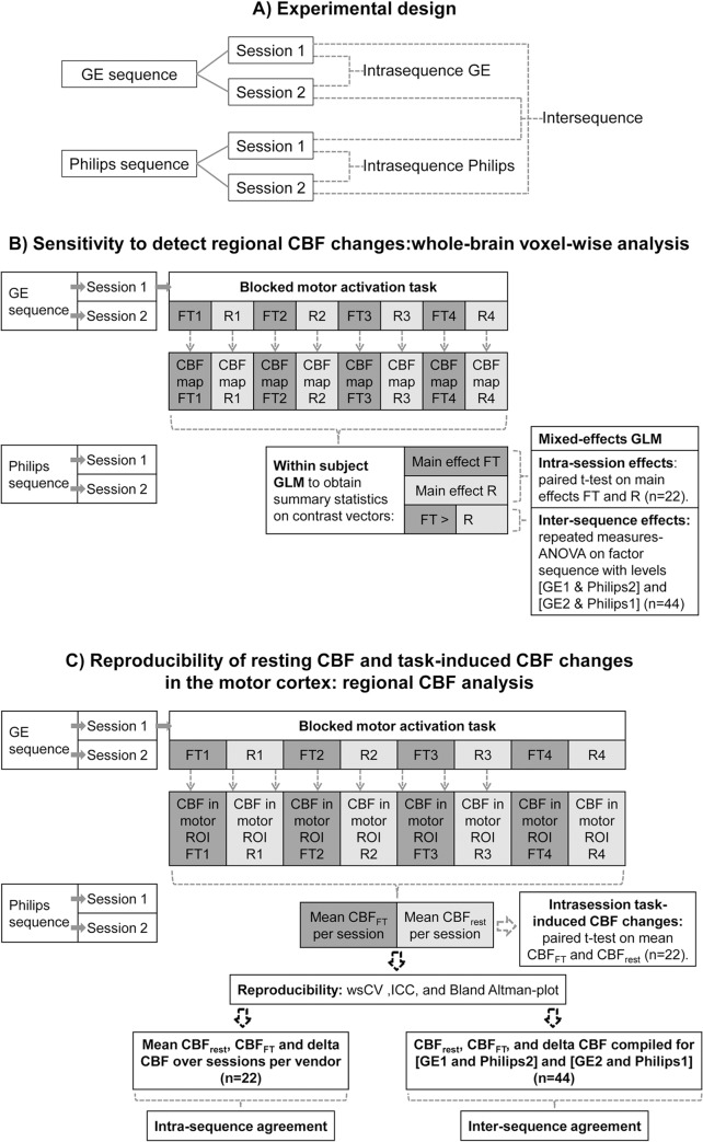
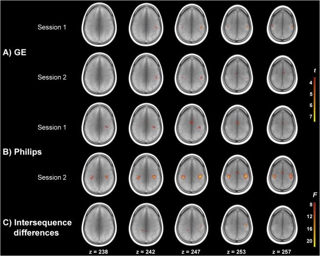
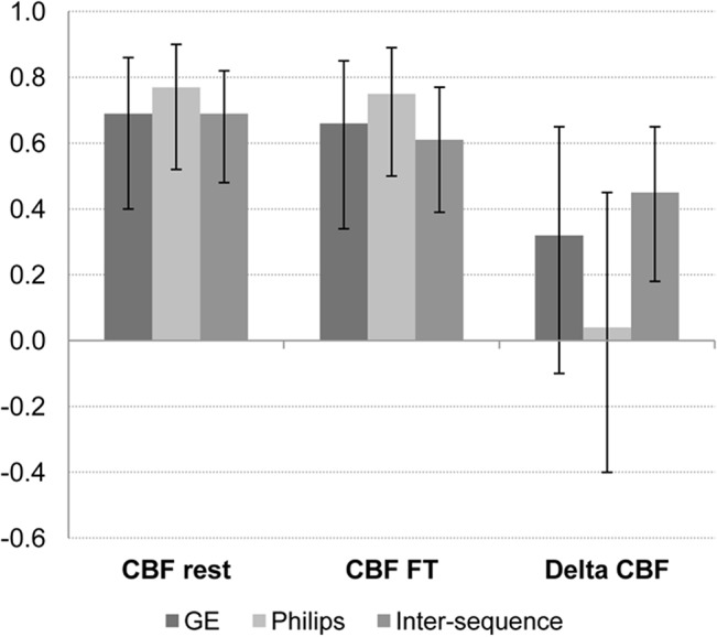
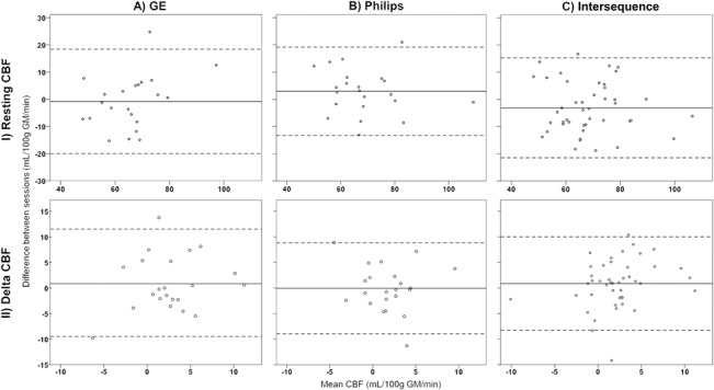
Arterial spin labeling (ASL) magnetic resonance imaging is increasingly used to quantify task-related brain activation. This study assessed functional ASL (fASL) using pseudo-continuous ASL (pCASL) product sequences from two vendors. By scanning healthy participants twice with each sequence while they performed a motor task, this study assessed functional ASL for 1) its sensitivity to detect task-related cerebral blood flow (CBF) changes, and 2) its reproducibility of resting CBF and absolute CBF changes (delta CBF) in the motor cortex. Whole-brain voxel-wise analyses showed that sensitivity for motor activation was sufficient with each sequence, and comparable between sequences. Reproducibility was assessed with within-subject coefficients of variation (wsCV) and intraclass correlation coefficients (ICC). Reproducibility of resting CBF was reasonably good within (wsCV: 14.1-15.7%; ICC: 0.69-0.77) and between sequences (wsCV: 15.1%; ICC: 0.69). Reproducibility of delta CBF was relatively low, both within (wsCV: 182-297%; ICC: 0.04-0.32) and between sequences (wsCV: 185%; ICC: 0.45), while inter-session variation was low. This may be due to delta CBF's small mean effect (0.77-1.32 mL/100g gray matter/min). In conclusion, fASL seems sufficiently sensitive to detect task-related changes on a group level, with acceptable inter-sequence differences. Resting CBF may provide a consistent baseline to compare task-related activation to, but absolute regional CBF changes are more variable, and should be interpreted cautiously when acquired with two pCASL product sequences.
Conflict of interest statement
Figures
Similar articles
-
Intra- and interscanner reliability and reproducibility of 3D whole-brain pseudo-continuous arterial spin-labeling MR perfusion at 3T.J Magn Reson Imaging. 2014 Feb;39(2):402-9. doi: 10.1002/jmri.24175. Epub 2013 May 30. J Magn Reson Imaging. 2014. PMID: 23723043
-
Test-retest reliability and reproducibility of long-label pseudo-continuous arterial spin labeling.Magn Reson Imaging. 2020 Nov;73:111-117. doi: 10.1016/j.mri.2020.07.010. Epub 2020 Jul 25. Magn Reson Imaging. 2020. PMID: 32717203
-
Multi-vendor reliability of arterial spin labeling perfusion MRI using a near-identical sequence: implications for multi-center studies.Neuroimage. 2015 Jun;113:143-52. doi: 10.1016/j.neuroimage.2015.03.043. Epub 2015 Mar 24. Neuroimage. 2015. PMID: 25818685
-
3D Pseudocontinuous arterial spin labeling in routine clinical practice: A review of clinically significant artifacts.J Magn Reson Imaging. 2016 Jan;43(1):11-27. doi: 10.1002/jmri.24873. Epub 2015 Apr 9. J Magn Reson Imaging. 2016. PMID: 25857715 Review.
-
Altered cerebral perfusion in bipolar disorder: A pCASL MRI study.Bipolar Disord. 2021 Mar;23(2):130-140. doi: 10.1111/bdi.12966. Epub 2020 Jul 25. Bipolar Disord. 2021. PMID: 32583570 Review.
Cited by
-
Three-dimensional arterial spin labeling imaging and dynamic susceptibility contrast perfusion-weighted imaging value in diagnosing glioma grade prior to surgery.Exp Ther Med. 2017 Jun;13(6):2691-2698. doi: 10.3892/etm.2017.4370. Epub 2017 Apr 20. Exp Ther Med. 2017. PMID: 28587332 Free PMC article.
-
Comparison of velocity-selective arterial spin labeling schemes.Magn Reson Med. 2021 Apr;85(4):2027-2039. doi: 10.1002/mrm.28572. Epub 2020 Oct 31. Magn Reson Med. 2021. PMID: 33128484 Free PMC article.
-
Test-retest reliability of cerebral blood flow in healthy individuals using arterial spin labeling: Findings from the EMBARC study.Magn Reson Imaging. 2018 Jan;45:26-33. doi: 10.1016/j.mri.2017.09.004. Epub 2017 Sep 6. Magn Reson Imaging. 2018. PMID: 28888770 Free PMC article.
-
A multicenter, single-arm, phase II clinical trial of adrenomedullin in patients with cerebral autosomal dominant arteriopathy with subcortical infarcts and leukoencephalopathy.Cereb Circ Cogn Behav. 2024 Feb 6;6:100211. doi: 10.1016/j.cccb.2024.100211. eCollection 2024. Cereb Circ Cogn Behav. 2024. PMID: 38375188 Free PMC article.
-
Comparison of test-retest reliability of BOLD and pCASL fMRI in a two-center study.BMC Med Imaging. 2022 Apr 3;22(1):62. doi: 10.1186/s12880-022-00791-9. BMC Med Imaging. 2022. PMID: 35366813 Free PMC article.
References
-
- Wang J, Aguirre GK, Kimberg DY, Roc AC, Li L, Detre JA. Arterial spin labeling perfusion fMRI with very low task frequency. Magn Reson Med. 2003;49: 796–802. - PubMed
Publication types
MeSH terms
Substances
LinkOut - more resources
Full Text Sources
Other Literature Sources
