. 2015 Jul 14;11(7):e1004386.
doi: 10.1371/journal.pcbi.1004386. eCollection 2015 Jul.

A Developmental Switch for Hebbian Plasticity

Affiliations

A Developmental Switch for Hebbian Plasticity

Marijn B Martens et al. PLoS Comput Biol. .

Abstract

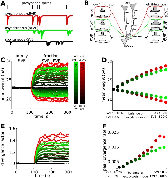
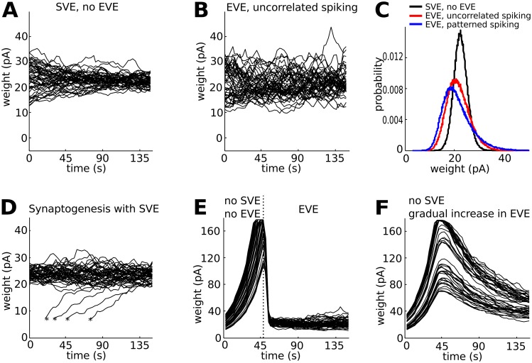
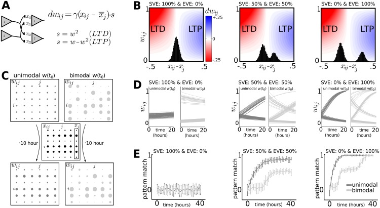
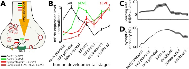
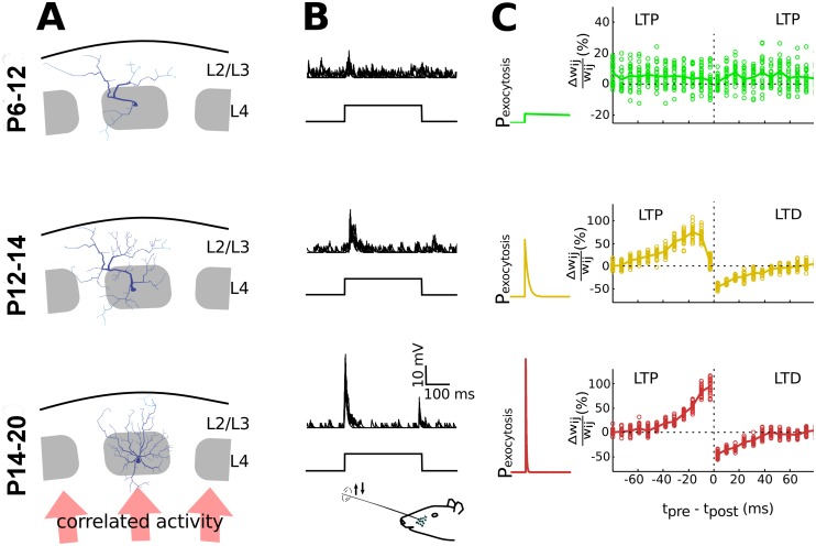
Hebbian forms of synaptic plasticity are required for the orderly development of sensory circuits in the brain and are powerful modulators of learning and memory in adulthood. During development, emergence of Hebbian plasticity leads to formation of functional circuits. By modeling the dynamics of neurotransmitter release during early postnatal cortical development we show that a developmentally regulated switch in vesicle exocytosis mode triggers associative (i.e. Hebbian) plasticity. Early in development spontaneous vesicle exocytosis (SVE), often considered as 'synaptic noise', is important for homogenization of synaptic weights and maintenance of synaptic weights in the appropriate dynamic range. Our results demonstrate that SVE has a permissive, whereas subsequent evoked vesicle exocytosis (EVE) has an instructive role in the expression of Hebbian plasticity. A timed onset for Hebbian plasticity can be achieved by switching from SVE to EVE and the balance between SVE and EVE can control the effective rate of Hebbian plasticity. We further show that this developmental switch in neurotransmitter release mode enables maturation of spike-timing dependent plasticity. A mis-timed or inadequate SVE to EVE switch may lead to malformation of brain networks thereby contributing to the etiology of neurodevelopmental disorders.


Conflict of interest statement

The authors have declared that no competing interests exist.

Figures

Fig 1
Fig 1. The balance between spontaneous and evoked vesicle exocytosis provides control for the rate of Hebbian plasticity.
We use the VTDP model represented by Eqs (1) to (8). A: Vesicle exocytosis can occur directly following a presynaptic spike (sEVE), with a high probability after a presynaptic spike (aEVE) or randomly, independent of a presynaptic spike (SVE). B: Each exocytosis mode can coordinate activity across the synapse. When EVE mode dominates the release process, there will be competition between synapses of neurons that have low (left presynaptic neuron) and high (right presynaptic neuron) firing rates, with the former decreasing in strength and the latter increasing in strength (top and middle synapses). There is no such competition for SVE dominated synapses (bottom synapses). C: Vesicle exocytosis is initially dominated by SVE, which maintains the synaptic strength distribution. Following a switch to EVE, the synapses at which many action potentials arrive (upper traces) are potentiated at the expense of synapses for the lower firing rate neurons (lower traces). The (partial) switch to EVE occurs at the dotted line and is either to aEVE or sEVE. The colorbar denotes the SVE-aEVE (green) and SVE-sEVE (red) balance. D: The final difference in weight for the synapses of the low- and high firing rate neurons depends on the SVE-EVE balance. Dots are the mean of the last 100 seconds for the upper (top) and lower (bottom) traces in panel C. Colors represent the same degree of balance as in panel C. E: The divergence factor between the synapses of the high and low firing rate neurons (upper and lower traces in panel C, respectively) increases after the SVE to EVE switch. F: The divergence rate is calculated as the change in divergence factor (panel E) per time unit. The maximal rate of divergence depends on the SVE-EVE balance, where a large fraction of EVE results in faster divergence. The divergence rate for equivalent strength is a bit lower for aEVE compared to sEVE. For purely SVE there is no divergence. The SVE-EVE balance can thus act to control the divergence rate during synaptic competition.
Fig 2
Fig 2. Additional functional roles of spontaneous vesicle exocytosis (SVE).
A: Synaptic weights that are initially heterogeneous, homogenize into a tight synaptic weight distribution in the case of SVE. Black lines are traces of 50 synaptic weights randomly selected from a network of 500 presynaptic neurons that connect to 10 postsynaptic neurons. The neurons fire uncorrelated action potentials at the same mean firing rate. B: Synaptic weights show large fluctuations in the case of EVE. As before, neurons fire uncorrelated action potentials at the same mean firing rate. C: Distribution of synaptic weights for SVE (black line), EVE with uncorrelated spiking activity (4.8 Hz, blue line) and EVE with patterned spiking activity (red line). For the patterned, a subset of the neurons (20%) have higher firing rates (8 Hz) compared to the others (4 Hz). Notice the broadening for uncorrelated and patterned spiking activity in the case of EVE. D: Synaptogenesis creates new synapses with small initial synaptic weights (denoted with a *). SVE incorporates the newly generated synapses into the homogeneous pool of all synaptic weights. E: In the absence of SVE, the synaptic weights increase due to the homeostatic mechanism until they reach the max synapse size. Synaptic weights are heterogeneous when the switch to evoked vesicle exocytosis (EVE) occurs. F: For a gradual increase of EVE, in the absence of SVE, the synaptic weights span a large dynamic range and are heterogeneous distributed.
Fig 3
Fig 3. The rate at which a pattern of action potentials is imprinted onto a configuration of synaptic weights depends on the initial weight distribution and the SVE-EVE balance.
We used the rate model described by Eqs (11) and (12). A: Presynaptic neurons i connect to postsynaptic neurons j, for which the synaptic weights are shaped by local competition between the vesicle exocytosis rates (xij relative to the average exocytosis rate onto the postsynaptic neuron (x¯ij)). The exocytosis rates are a combination of SVE (randomly distributed across synapses) and EVE (strong for the subset of highly active presynaptic neurons). B: The degree of potentiation (blue, positive rate of change in dwij) and depression (red, negative rate of change) depends on whether the relative exocytosis rate is bimodal versus unimodal (black histograms). C: The initial synaptic weights (w) have either a unimodal distribution (left) or a bimodal distribution (right). A pattern of activity (x) is presented to both networks. Using the rate model, we can clearly see the activity pattern is represented in the weights of the network that started with the unimodal distribution, but not for the initial bimodal distribution. Dot size represents weight (w ij) or exocytosis rate (x ij). D: The rate at which synaptic weights converge to a configuration corresponding to the applied patterns of action potentials depends on the SVE-EVE balance. Dark and light gray traces start from an initial unimodal and bimodal distribution respectively. E: Matching the synaptic weights to the pattern of action-potential activity (pattern match, Eq (13)) takes significantly longer when the initial weights have a bimodal distribution.
Fig 4
Fig 4. During cortical development there is a switch from exocytosis dominated by spontaneous exocytosis to asynchronous and synchronous vesicle exocytosis.
A: The SVE-EVE balance is regulated by Ca2+ sensing proteins (Doc2s and synaptotagmins). Complexin1 promotes Synaptotagmin1 mediated synchronous exocytosis while blocking Spontaneous and asynchronous exocytosis [29]. B: In humans, the relative mRNA expression of these sensors varies in time. Early in development there are high levels of Doc2b (SVE, black line), which decay during the intermediate developmental stage at which time there is an increase in Doc2a (aEVE, green line) expression. The expression of Synaptotagmin1/Complexin1 (sEVE, red lines) increases throughout neurodevelopment, reaching their maximal expression levels during late development [31]. C: By implementing the mRNA expression levels as ratios for SVE, aEVE and sEVE, a period of rapid learning is observed early in development during which SVE decreases and aEVE and sEVE increase. During this period the wiring in the neural circuitry is formed, after which the learning rate decreases. Gray shade is standard deviation. D: During early developmental periods synaptogenesis is prominent, until activity-dependent pruning reduces the synaptic density later in life.
Fig 5
Fig 5. Predictions for the role of the SVE to EVE switch in refinement of cortical neural circuits.
A: During early development arborization of barrel cortical neurons is sparse, and does not respect the columnar boundaries. Neurite outgrowth occurs during the intermediate developmental period and in late development, as synapses mature, the extensive arborizations outside the column are pruned in an activity dependent manner (illustration based on the results from [1], using the Trees Toolbox [37]). B: Whisker deflection induces synaptic responses in layer 2/3 of the barrel cortex. The input to L2/3 is largely uncorrelated at P12, with a rapid switch to stimulus-driven responses during development [2]. At P12-P14 the stimulus-induced activity is typically prolonged and with high variability, whereas at a later stage (P20) the responses are more precisely time-locked to the stimulus [2]. C: By modeling changes in the release probability from SVE to highly time-locked release during development, Hebbian plasticity behavior changes from an immature to mature STDP rule. The maturation process of the STDP rule is described in vitro for layer 4 –layer 2/3 synapses during these critical developmental stages [19]. The calcium time constant (Eq (4)) was 1 second (top), 7 millisecond (middle) and 2 millisecond (bottom), and 50 stimuli were given for each tpre - tpost bin.

Similar articles

Cited by

References

    1. Bender K, Rangel J, Feldman D. Development of Columnar Topography in the Excitatory Layer 4 to Layer 2/3 Projection in Rat Barrel Cortex. J Neurosci. 2003;23(25):8759–70. - PMC - PubMed
    1. Stern E, Maravall M, Svoboda K. Rapid Development and Plasticity of Layer 2/3 Maps in Rat Barrel Cortex In Vivo. Neuron. 2002;31:305–15. - PubMed
    1. Narboux-Nûme N, Evrard A, Ferezou I, Erzurumlu R, Kaeser P, Lainú J, et al. Neurotransmitter Release at the Thalamocortical Synapse Instructs Barrel Formation But Not Axon Patterning in the Somatosensory Cortex. J Neurosci. 2012;32(18):6183–96. 10.1523/JNEUROSCI.0343-12.2012 - DOI - PMC - PubMed
    1. Cohen-Cory S. The Developing Synapse: Construction and Modulation of Synaptic Structures and Circuits. Science. 2002;298(5594):770–6. - PubMed
    1. Ko H, Cossell L, Baragli C, Antolik J, Clopath C, Hofer S, et al. The emergence of functional microcircuits in visual cortex. Nature. 2013;496(7443):96–100. 10.1038/nature12015 - DOI - PMC - PubMed

Publication types

Substances